 Movie
Movie Controller


+ Open data

- Basic information
| Entry | Database: PDB / ID: 7jl7 | ||||||
|---|---|---|---|---|---|---|---|
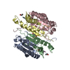
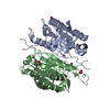
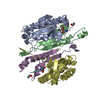
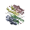
| Title | Zebrafish Caspase N213T | ||||||
 Components Components |
| ||||||
 Keywords Keywords |  CELL CYCLE / enzyme specificity / apoptosis / caspase / zebrafish / protein evolution CELL CYCLE / enzyme specificity / apoptosis / caspase / zebrafish / protein evolution | ||||||
| Function / homology |  Function and homology information Function and homology informationregulation of collateral sprouting in absence of injury / Activation of caspases through apoptosome-mediated cleavage / Apoptotic cleavage of cellular proteins / SMAC, XIAP-regulated apoptotic response / Apoptosis induced DNA fragmentation / Signaling by Hippo / Caspase-mediated cleavage of cytoskeletal proteins / : / Caspase activation via Dependence Receptors in the absence of ligand / Other interleukin signaling ...regulation of collateral sprouting in absence of injury / Activation of caspases through apoptosome-mediated cleavage / Apoptotic cleavage of cellular proteins / SMAC, XIAP-regulated apoptotic response / Apoptosis induced DNA fragmentation / Signaling by Hippo / Caspase-mediated cleavage of cytoskeletal proteins / : / Caspase activation via Dependence Receptors in the absence of ligand / Other interleukin signaling / : / Pyroptosis / : / : / Apoptotic cleavage of cell adhesion proteins / Regulation of TNFR1 signaling / cysteine-type endopeptidase activity involved in apoptotic process / cellular response to antibiotic / cellular response to retinoic acid / keratinocyte differentiation / erythrocyte differentiation / neuron differentiation / cellular response to xenobiotic stimulus / peptidase activity / positive regulation of apoptotic process / apoptotic process / proteolysisSimilarity search - Function | ||||||
| Biological species |  Danio rerio (zebrafish) Danio rerio (zebrafish)synthetic construct (others) | ||||||
| Method | X-RAY DIFFRACTION / SYNCHROTRON / MOLECULAR REPLACEMENT / Resolution: 2.05 Å | ||||||
 Authors Authors | Clark, A.C. / Swartz, P.D. | ||||||
| Funding support |  United States, 1items United States, 1items
| ||||||
 Citation Citation | Journal: Biosci.Rep. / Year: 2021 Title: Remodeling hydrogen bond interactions results in relaxed specificity of Caspase-3. Authors: Yao, L. / Swartz, P. / Hamilton, P.T. / Clark, A.C. | ||||||
| History |
|
- Structure visualization
| Structure viewer | Molecule: MolmilJmol/JSmol |
|---|
- Downloads & links
-Download
| PDBx/mmCIF format | 7jl7.cif.gz | 110.4 KB | Display | PDBx/mmCIF format |
|---|---|---|---|---|
| PDB format | pdb7jl7.ent.gz | 82.6 KB | Display | PDB format |
| PDBx/mmJSON format | 7jl7.json.gz | Tree view | PDBx/mmJSON format | |
| Others |  Other downloads Other downloads |
-Validation report
| Arichive directory | https://data.pdbj.org/pub/pdb/validation_reports/jl/7jl7ftp://data.pdbj.org/pub/pdb/validation_reports/jl/7jl7 | HTTPS FTP |
|---|
-Related structure data
| Related structure data |  5jftS S: Starting model for refinement |
|---|---|
| Similar structure data |


-Links
 PDBj
PDBj

- Assembly
| Deposited unit | 
| ||||||||
|---|---|---|---|---|---|---|---|---|---|
| 1 |
| ||||||||
| Unit cell |
|
-Components
| #1: Protein | / Caspase-3 Mass: 19512.988 Da / Num. of mol.: 2 Source method: isolated from a genetically manipulated source Source: (gene. exp.) Danio rerio (zebrafish) / Gene: casp3a, casp3 / Production host:  Escherichia coli (E. coli) / References: UniProt: Q98UI8, UniProt: B8JK21*PLUS Escherichia coli (E. coli) / References: UniProt: Q98UI8, UniProt: B8JK21*PLUS#2: Protein | / Caspase-3Mass: 12033.823 Da / Num. of mol.: 2 / Mutation: N213T Source method: isolated from a genetically manipulated source Source: (gene. exp.) Danio rerio (zebrafish) / Gene: casp3a, casp3 / Production host: Escherichia coli (E. coli) / References: UniProt: Q98UI8#3: Protein/peptide | | Mass: 476.435 Da / Num. of mol.: 1 / Source method: obtained synthetically / Source: (synth.) synthetic construct (others) #4: Water | ChemComp-HOH / | Water Mass: 18.015 Da / Num. of mol.: 109 / Source method: isolated from a natural source / Formula: H2O Mass: 18.015 Da / Num. of mol.: 109 / Source method: isolated from a natural source / Formula: H2O |
|---|
-Experimental details
-Experiment
| Experiment | Method: X-RAY DIFFRACTION / Number of used crystals: 1 |
|---|
- Sample preparation
| Crystal | Density Matthews: 2.13 Å3/Da / Density % sol: 42.35 % |
|---|---|
| Crystal grow | Temperature: 291 K / Method: vapor diffusion, hanging drop Details: Protein was dialyzed in a buffer of 10 mM Tris-HCl, pH 8.5, 1 mM DTT, concentrated to 4 mg/mL, and inhibitor Ac-DEVD-CHO was added at a 5:1 (w/w) inhibitor/protein ratio. Crystals were ...Details: Protein was dialyzed in a buffer of 10 mM Tris-HCl, pH 8.5, 1 mM DTT, concentrated to 4 mg/mL, and inhibitor Ac-DEVD-CHO was added at a 5:1 (w/w) inhibitor/protein ratio. Crystals were obtained at 18 C by the hanging drop vapor diffusion method using a 4 uL drop that contained equal amounts of protein and reservoir solution. Each well contained a reservoir solution (500 uL) of 100 mM sodium citrate, pH 5.4, 23% PEG 6000, 10 mM DTT, and 3 mM NaN3. Flat, sheet-like crystals appeared within 14 days, and we used microseeding to obtain diffraction-quality crystals. In this case, crystal trays were set up as described above and incubated for 24 hours. The flat sheet crystals were collected and treated with seed beads using a kit from Hampton Research. A 4 uL drop containing flat sheet crystals was added to a tube containing seed beads. Reservoir solution (10 uL) was pipetted on the cover slide to remove all crystals from the coverslip, and the procedure was repeated five times. The resulting mixture of crystals and seed beads was vortexed for 30 seconds and cooled on ice for 10 seconds, and the procedure was repeated six times. Serial dilutions of the treated crystals were set up from 10-1 to 10-3, and 0.5 uL of the 10-2 dilution crystal seeds was added into the drops of the 24-hour crystal tray. Larger cube-shaped crystals appeared within 14 days. The crystals were collected and frozen in liquid nitrogen following the addition of 20% MPD plus the reservoir solution. |
-Data collection
| Diffraction | Mean temperature: 100 K / Ambient temp details: Constant temperature / Serial crystal experiment: N |
|---|---|
| Diffraction source | Source: SYNCHROTRON / Site: APS / Beamline: 22-BM / Wavelength: 1 Å |
| Detector | Type: MARMOSAIC 300 mm CCD / Detector: CCD / Date: Aug 18, 2018 |
| Radiation | Monochromator: Insertion device / Protocol: SINGLE WAVELENGTH / Monochromatic (M) / Laue (L): M / Scattering type: x-ray |
| Radiation wavelength | Wavelength: 1 Å / Relative weight: 1 |
| Reflection | Resolution: 2.05→35.91 Å / Num. obs: 32428 / % possible obs: 90.51 % / Redundancy: 5.1 % / CC1/2: 0.843 / Net I/σ(I): 30.77 |
| Reflection shell | Resolution: 2.07→2.11 Å / Num. unique obs: 91 / CC1/2: 0.069 |
- Processing
| Software |
| |||||||||||||||||||||||||||||||||||||||||||||||||||||||||||||||||||||||||||||||||||||||||||||||||||||||||
|---|---|---|---|---|---|---|---|---|---|---|---|---|---|---|---|---|---|---|---|---|---|---|---|---|---|---|---|---|---|---|---|---|---|---|---|---|---|---|---|---|---|---|---|---|---|---|---|---|---|---|---|---|---|---|---|---|---|---|---|---|---|---|---|---|---|---|---|---|---|---|---|---|---|---|---|---|---|---|---|---|---|---|---|---|---|---|---|---|---|---|---|---|---|---|---|---|---|---|---|---|---|---|---|---|---|---|
| Refinement | Method to determine structure: MOLECULAR REPLACEMENT Starting model: 5JFT Resolution: 2.05→35.91 Å / SU ML: 0.31 / Cross valid method: THROUGHOUT / σ(F): 1.33 / Phase error: 28.06 / Stereochemistry target values: ML
| |||||||||||||||||||||||||||||||||||||||||||||||||||||||||||||||||||||||||||||||||||||||||||||||||||||||||
| Solvent computation | Shrinkage radii: 0.9 Å / VDW probe radii: 1.11 Å / Solvent model: FLAT BULK SOLVENT MODEL | |||||||||||||||||||||||||||||||||||||||||||||||||||||||||||||||||||||||||||||||||||||||||||||||||||||||||
| Displacement parameters | Biso max: 99.05 Å2 / Biso mean: 37.4882 Å2 / Biso min: 16.64 Å2 | |||||||||||||||||||||||||||||||||||||||||||||||||||||||||||||||||||||||||||||||||||||||||||||||||||||||||
| Refinement step | Cycle: final / Resolution: 2.05→35.91 Å
| |||||||||||||||||||||||||||||||||||||||||||||||||||||||||||||||||||||||||||||||||||||||||||||||||||||||||
| LS refinement shell | Refine-ID: X-RAY DIFFRACTION / Rfactor Rfree error: 0 / Total num. of bins used: 14
|
