 Movie
Movie Controller


+ Open data

- Basic information
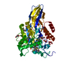
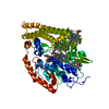
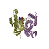
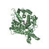
| Entry | Database: PDB / ID: 5k16 | ||||||
|---|---|---|---|---|---|---|---|
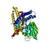
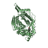
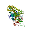
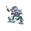
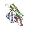
| Title | Crystal structure of free Ubiquitin-specific protease 12 | ||||||
 Components Components | Ubiquitin carboxyl-terminal hydrolase 12 | ||||||
 Keywords Keywords |  HYDROLASE / deubiquitination / deubiquitnating enzyme / DUB HYDROLASE / deubiquitination / deubiquitnating enzyme / DUB | ||||||
| Function / homology |  Function and homology information Function and homology informationprotein deubiquitination / ubiquitinyl hydrolase 1 / cysteine-type deubiquitinase activity / Ub-specific processing proteases / cysteine-type endopeptidase activity / proteolysis / nucleoplasm / metal ion binding / nucleus / plasma membrane ...protein deubiquitination / ubiquitinyl hydrolase 1 / cysteine-type deubiquitinase activity / Ub-specific processing proteases / cysteine-type endopeptidase activity / proteolysis / nucleoplasm / metal ion binding / nucleus / plasma membrane / cytosol / cytoplasmSimilarity search - Function | ||||||
| Biological species |  Homo sapiens (human) Homo sapiens (human) | ||||||
| Method | X-RAY DIFFRACTION / SYNCHROTRON / MOLECULAR REPLACEMENT / Resolution: 2.599 Å | ||||||
 Authors Authors | Li, H. / D'Andrea, A.D. / Zheng, N. | ||||||
| Funding support |  United States, 1items United States, 1items
| ||||||
 Citation Citation | Journal: Mol.Cell / Year: 2016 Title: Allosteric Activation of Ubiquitin-Specific Proteases by beta-Propeller Proteins UAF1 and WDR20. Authors: Li, H. / Lim, K.S. / Kim, H. / Hinds, T.R. / Jo, U. / Mao, H. / Weller, C.E. / Sun, J. / Chatterjee, C. / D'Andrea, A.D. / Zheng, N. | ||||||
| History |
|
- Structure visualization
| Structure viewer | Molecule: MolmilJmol/JSmol |
|---|
- Downloads & links
-Download
| PDBx/mmCIF format | 5k16.cif.gz | 144.9 KB | Display | PDBx/mmCIF format |
|---|---|---|---|---|
| PDB format | pdb5k16.ent.gz | 110.6 KB | Display | PDB format |
| PDBx/mmJSON format | 5k16.json.gz | Tree view | PDBx/mmJSON format | |
| Others |  Other downloads Other downloads |
-Validation report
| Arichive directory | https://data.pdbj.org/pub/pdb/validation_reports/k1/5k16ftp://data.pdbj.org/pub/pdb/validation_reports/k1/5k16 | HTTPS FTP |
|---|
-Related structure data
| Related structure data |  5k19C  5k1aC  5k1bC 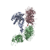 5k1cC 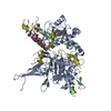 3m99S C: citing same article ( S: Starting model for refinement |
|---|---|
| Similar structure data |

-Links
 PDBj
PDBj- Assembly
| Deposited unit | 
| ||||||||
|---|---|---|---|---|---|---|---|---|---|
| 1 | 
| ||||||||
| 2 | 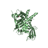
| ||||||||
| 3 |
| ||||||||
| Unit cell |
| ||||||||
| Details | Monomer as determined by size-exclusion chromatography |
-Components
| #1: Protein | Mass: 41256.684 Da / Num. of mol.: 2 / Fragment: residues 16-370 Source method: isolated from a genetically manipulated source Source: (gene. exp.) Homo sapiens (human) / Gene: USP12, UBH1, USP12L1 / Production host:  Escherichia coli K-12 (bacteria) / References: UniProt: O75317, ubiquitinyl hydrolase 1 Escherichia coli K-12 (bacteria) / References: UniProt: O75317, ubiquitinyl hydrolase 1#2: Chemical |   Mass: 65.409 Da / Num. of mol.: 2 / Source method: obtained synthetically / Formula: Zn Mass: 65.409 Da / Num. of mol.: 2 / Source method: obtained synthetically / Formula: Zn#3: Chemical | Glycerol  Mass: 92.094 Da / Num. of mol.: 2 / Source method: obtained synthetically / Formula: C3H8O3 Mass: 92.094 Da / Num. of mol.: 2 / Source method: obtained synthetically / Formula: C3H8O3#4: Water | ChemComp-HOH / | Water Mass: 18.015 Da / Num. of mol.: 56 / Source method: isolated from a natural source / Formula: H2O Mass: 18.015 Da / Num. of mol.: 56 / Source method: isolated from a natural source / Formula: H2O |
|---|
-Experimental details
-Experiment
| Experiment | Method: X-RAY DIFFRACTION / Number of used crystals: 1 |
|---|
- Sample preparation
| Crystal | Density Matthews: 2.52 Å3/Da / Density % sol: 51.24 % |
|---|---|
| Crystal grow | Temperature: 277 K / Method: vapor diffusion, hanging drop / pH: 7 Details: 0.1 M Tris-HCl, 18% PEG 3350, 0.2 M Lithium Sulfate |
-Data collection
| Diffraction | Mean temperature: 100 K |
|---|---|
| Diffraction source | Source: SYNCHROTRON / Site: ALS / Beamline: 8.2.1 / Wavelength: 1 Å |
| Detector | Type: ADSC QUANTUM 315 / Detector: CCD / Date: May 15, 2013 |
| Radiation | Protocol: SINGLE WAVELENGTH / Monochromatic (M) / Laue (L): M / Scattering type: x-ray |
| Radiation wavelength | Wavelength: 1 Å / Relative weight: 1 |
| Reflection | Resolution: 2.599→50 Å / Num. obs: 24515 / % possible obs: 99.9 % / Redundancy: 4 % / Rmerge(I) obs: 0.091 / Rsym value: 0.105 / Net I/σ(I): 14.3 |
| Reflection shell | Resolution: 2.599→2.64 Å / Redundancy: 4 % / Rmerge(I) obs: 0.74 / Mean I/σ(I) obs: 1.4 / % possible all: 99.9 |
- Processing
| Software |
| ||||||||||||||||||||||||||||||||||||||||||||||||||||||||||||||||||||||
|---|---|---|---|---|---|---|---|---|---|---|---|---|---|---|---|---|---|---|---|---|---|---|---|---|---|---|---|---|---|---|---|---|---|---|---|---|---|---|---|---|---|---|---|---|---|---|---|---|---|---|---|---|---|---|---|---|---|---|---|---|---|---|---|---|---|---|---|---|---|---|---|
| Refinement | Method to determine structure: MOLECULAR REPLACEMENT Starting model: 3M99 Resolution: 2.599→42.451 Å / SU ML: 0.36 / Cross valid method: FREE R-VALUE / σ(F): 1.35 / Phase error: 25.03 / Stereochemistry target values: ML
| ||||||||||||||||||||||||||||||||||||||||||||||||||||||||||||||||||||||
| Solvent computation | Shrinkage radii: 0.9 Å / VDW probe radii: 1.11 Å / Solvent model: FLAT BULK SOLVENT MODEL | ||||||||||||||||||||||||||||||||||||||||||||||||||||||||||||||||||||||
| Refinement step | Cycle: LAST / Resolution: 2.599→42.451 Å
| ||||||||||||||||||||||||||||||||||||||||||||||||||||||||||||||||||||||
| Refine LS restraints |
| ||||||||||||||||||||||||||||||||||||||||||||||||||||||||||||||||||||||
| LS refinement shell |
|
