 Movie
Movie Controller

[English] 日本語
 Yorodumi
- PDB-2e81: Cytochrome c Nitrite Reductase from Wolinella succinogenes with b... -
+ Open data

- Basic information
| Entry | Database: PDB / ID: 2.0E+81 | ||||||
|---|---|---|---|---|---|---|---|
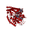
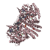
| Title | Cytochrome c Nitrite Reductase from Wolinella succinogenes with bound intermediate hydroxylamine | ||||||
 Components Components | Cytochrome c-552 | ||||||
 Keywords Keywords |  OXIDOREDUCTASE / multiheme cytochrome / nitrite reductase / reaction intermediate OXIDOREDUCTASE / multiheme cytochrome / nitrite reductase / reaction intermediate | ||||||
| Function / homology |  Function and homology informationnitrite reductase (cytochrome; ammonia-forming) / nitrite reductase (cytochrome, ammonia-forming) activity / : / nitrate assimilation / outer membrane-bounded periplasmic space / calcium ion binding / heme binding Function and homology informationnitrite reductase (cytochrome; ammonia-forming) / nitrite reductase (cytochrome, ammonia-forming) activity / : / nitrate assimilation / outer membrane-bounded periplasmic space / calcium ion binding / heme bindingSimilarity search - Function | ||||||
| Biological species |  Wolinella succinogenes (bacteria) Wolinella succinogenes (bacteria) | ||||||
| Method | X-RAY DIFFRACTION / SYNCHROTRON / MOLECULAR REPLACEMENT / Resolution: 2 Å | ||||||
 Authors Authors | Einsle, O. / Kroneck, P.M.H. | ||||||
 Citation Citation | Journal: J.Am.Chem.Soc. / Year: 2002 Title: Mechanism of the six-electron reduction of nitrite to ammonia by cytochrome c nitrite reductase Authors: Einsle, O. / Messerschmidt, A. / Huber, R. / Kroneck, P.M.H. / Neese, F. #1: Journal: Nature / Year: 1999Title: Structure of cytochrome c nitrite reductase Authors: Einsle, O. / Messerschmidt, A. / Stach, P. / Bourenkov, G.P. / Bartunik, H.D. / Huber, R. / Kroneck, P.M.H. #2: Journal: J.Biol.Chem. / Year: 2000Title: Cytochrome c nitrite reductase from Wolinella succinogenes. Structure at 1.6 A resolution, inhibitor binding, and heme-packing motifs Authors: Einsle, O. / Stach, P. / Messerschmidt, A. / Simon, J. / Kroeger, A. / Huber, R. / Kroneck, P.M.H. | ||||||
| History |
|
- Structure visualization
| Structure viewer | Molecule: MolmilJmol/JSmol |
|---|
- Downloads & links
-Download
| PDBx/mmCIF format | 2e81.cif.gz | 127.2 KB | Display | PDBx/mmCIF format |
|---|---|---|---|---|
| PDB format | pdb2e81.ent.gz | 97.1 KB | Display | PDB format |
| PDBx/mmJSON format | 2e81.json.gz | Tree view | PDBx/mmJSON format | |
| Others |  Other downloads Other downloads |
-Validation report
| Arichive directory | https://data.pdbj.org/pub/pdb/validation_reports/e8/2e81ftp://data.pdbj.org/pub/pdb/validation_reports/e8/2e81 | HTTPS FTP |
|---|
-Related structure data
| Related structure data |  2e80C  1fs7S S: Starting model for refinement C: citing same article ( |
|---|---|
| Similar structure data |


-Links
 PDBj
PDBj


- Assembly
| Deposited unit | 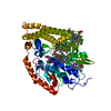
| ||||||||
|---|---|---|---|---|---|---|---|---|---|
| 1 | 
| ||||||||
| Unit cell |
| ||||||||
| Components on special symmetry positions |
| ||||||||
| Details | Biological unit is a dimer, created by the crystallographic twofold axis. |
-Components
-Protein , 1 types, 1 molecules A
| #1: Protein | Mass: 55343.602 Da / Num. of mol.: 1 / Source method: isolated from a natural source / Source: (natural) Wolinella succinogenes (bacteria) / Strain: DSM 1740References: UniProt: Q9S1E5, nitrite reductase (cytochrome; ammonia-forming) |
|---|
-Non-polymers , 5 types, 441 molecules 


| #2: Chemical | ChemComp-CA /  Mass: 40.078 Da / Num. of mol.: 1 / Source method: obtained synthetically / Formula: Ca Mass: 40.078 Da / Num. of mol.: 1 / Source method: obtained synthetically / Formula: Ca | ||||||
|---|---|---|---|---|---|---|---|
| #3: Chemical | Yttrium Mass: 88.906 Da / Num. of mol.: 3 / Source method: obtained synthetically / Formula: Y Mass: 88.906 Da / Num. of mol.: 3 / Source method: obtained synthetically / Formula: Y#4: Chemical | ChemComp-HEM / Heme B Mass: 616.487 Da / Num. of mol.: 5 / Source method: obtained synthetically / Formula: C34H32FeN4O4 Mass: 616.487 Da / Num. of mol.: 5 / Source method: obtained synthetically / Formula: C34H32FeN4O4#5: Chemical | ChemComp-HOA / | Hydroxylamine Mass: 33.030 Da / Num. of mol.: 1 / Source method: obtained synthetically / Formula: H3NO Mass: 33.030 Da / Num. of mol.: 1 / Source method: obtained synthetically / Formula: H3NO#6: Water | ChemComp-HOH / | WaterMass: 18.015 Da / Num. of mol.: 431 / Source method: isolated from a natural source / Formula: H2O |
-Experimental details
-Experiment
| Experiment | Method: X-RAY DIFFRACTION / Number of used crystals: 1 |
|---|
- Sample preparation
| Crystal | Density Matthews: 3.02 Å3/Da / Density % sol: 59.3 % |
|---|---|
| Crystal grow | Temperature: 293 K / Method: vapor diffusion, sitting drop / pH: 5.7 Details: 12% PEG 4000, 0.2M ammonium sulfate, 0.015M yttrium chloride, 0.1M sodium acetate, pH 5.7, VAPOR DIFFUSION, SITTING DROP, temperature 293K |
-Data collection
| Diffraction | Mean temperature: 100 K |
|---|---|
| Diffraction source | Source: SYNCHROTRON / Site: MPG/DESY, HAMBURG  / Beamline: BW6 / Wavelength: 1.05 Å / Beamline: BW6 / Wavelength: 1.05 Å |
| Detector | Type: MAR CCD 165 mm / Detector: CCD / Date: Feb 10, 2001 / Details: mirrors |
| Radiation | Monochromator: graphite / Protocol: SINGLE WAVELENGTH / Monochromatic (M) / Laue (L): M / Scattering type: x-ray |
| Radiation wavelength | Wavelength: 1.05 Å / Relative weight: 1 |
| Reflection | Resolution: 2→25 Å / Num. all: 46010 / Num. obs: 45781 / % possible obs: 99.5 % / Observed criterion σ(I): 2 / Biso Wilson estimate: 38.5 Å2 / Rmerge(I) obs: 0.079 / Net I/σ(I): 12.8 |
| Reflection shell | Resolution: 2→2.1 Å / Rmerge(I) obs: 0.488 / Mean I/σ(I) obs: 2 / % possible all: 99.7 |
- Processing
| Software |
| |||||||||||||||||||||||||
|---|---|---|---|---|---|---|---|---|---|---|---|---|---|---|---|---|---|---|---|---|---|---|---|---|---|---|
| Refinement | Method to determine structure: MOLECULAR REPLACEMENT Starting model: 1FS7 Resolution: 2→25 Å / Isotropic thermal model: isotropic / Cross valid method: THROUGHOUT / σ(F): 2 / Stereochemistry target values: Engh & Huber
| |||||||||||||||||||||||||
| Displacement parameters | Biso mean: 38.5 Å2
| |||||||||||||||||||||||||
| Refinement step | Cycle: LAST / Resolution: 2→25 Å
| |||||||||||||||||||||||||
| Refine LS restraints |
|
