 Movie
Movie Controller

[English] 日本語
 Yorodumi
- PDB-6g8v: Crystal structure of the light chain of botulinum neurotoxin X (r... -
+ Open data

- Basic information
| Entry | Database: PDB / ID: 6g8v | ||||||
|---|---|---|---|---|---|---|---|
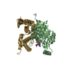
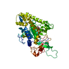
| Title | Crystal structure of the light chain of botulinum neurotoxin X (residues 2-442) | ||||||
 Components Components | Light chain of botulinum neurotoxin X (res. 2-442) | ||||||
 Keywords Keywords |  TOXIN / neurotoxin / endopeptidase TOXIN / neurotoxin / endopeptidase | ||||||
| Function / homology |  Function and homology informationbontoxilysin / host cell presynaptic membrane / host cell cytoplasmic vesicle / host cell cytosol / protein transmembrane transporter activity / metalloendopeptidase activity / toxin activity / host cell plasma membrane / proteolysis / zinc ion binding ...bontoxilysin / host cell presynaptic membrane / host cell cytoplasmic vesicle / host cell cytosol / protein transmembrane transporter activity / metalloendopeptidase activity / toxin activity / host cell plasma membrane / proteolysis / zinc ion binding / extracellular region / membrane Function and homology informationbontoxilysin / host cell presynaptic membrane / host cell cytoplasmic vesicle / host cell cytosol / protein transmembrane transporter activity / metalloendopeptidase activity / toxin activity / host cell plasma membrane / proteolysis / zinc ion binding ...bontoxilysin / host cell presynaptic membrane / host cell cytoplasmic vesicle / host cell cytosol / protein transmembrane transporter activity / metalloendopeptidase activity / toxin activity / host cell plasma membrane / proteolysis / zinc ion binding / extracellular region / membraneSimilarity search - Function | ||||||
| Biological species |   Clostridium botulinum (bacteria) Clostridium botulinum (bacteria) | ||||||
| Method | X-RAY DIFFRACTION / SYNCHROTRON / MOLECULAR REPLACEMENT / Resolution: 1.45 Å | ||||||
 Authors Authors | Wu, Y. / Kammerer, R. | ||||||
| Funding support |  Switzerland, 1items Switzerland, 1items
| ||||||
 Citation Citation | Journal: To Be Published Title: Crystal structure of the light chain of botulinum neurotoxin X (residues 2-442) Authors: Wu, Y. / Kammerer, R. | ||||||
| History |
|
- Structure visualization
| Structure viewer | Molecule: MolmilJmol/JSmol |
|---|
- Downloads & links
-Download
| PDBx/mmCIF format | 6g8v.cif.gz | 270.4 KB | Display | PDBx/mmCIF format |
|---|---|---|---|---|
| PDB format | pdb6g8v.ent.gz | 220.1 KB | Display | PDB format |
| PDBx/mmJSON format | 6g8v.json.gz | Tree view | PDBx/mmJSON format | |
| Others |  Other downloads Other downloads |
-Validation report
| Arichive directory | https://data.pdbj.org/pub/pdb/validation_reports/g8/6g8vftp://data.pdbj.org/pub/pdb/validation_reports/g8/6g8v | HTTPS FTP |
|---|
-Related structure data
| Related structure data | 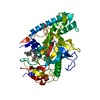 3qizS S: Starting model for refinement |
|---|---|
| Similar structure data |


-Links
 PDBj
PDBj- Assembly
| Deposited unit | 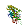
| ||||||||
|---|---|---|---|---|---|---|---|---|---|
| 1 |
| ||||||||
| Unit cell |
|
-Components
| #1: Protein | Mass: 52174.859 Da / Num. of mol.: 1 Source method: isolated from a genetically manipulated source Source: (gene. exp.) Clostridium botulinum (bacteria) / Production host: Escherichia coli BL21(DE3) (bacteria) / References: UniProt: P0DPK1*PLUS | ||
|---|---|---|---|
| #2: Chemical | ChemComp-ZN /   Mass: 65.409 Da / Num. of mol.: 1 / Source method: obtained synthetically / Formula: Zn Mass: 65.409 Da / Num. of mol.: 1 / Source method: obtained synthetically / Formula: Zn | ||
| #3: Chemical | ChemComp-GOL / Glycerol  Mass: 92.094 Da / Num. of mol.: 4 / Source method: obtained synthetically / Formula: C3H8O3 Mass: 92.094 Da / Num. of mol.: 4 / Source method: obtained synthetically / Formula: C3H8O3#4: Water | ChemComp-HOH / | Water Mass: 18.015 Da / Num. of mol.: 435 / Source method: isolated from a natural source / Formula: H2O Mass: 18.015 Da / Num. of mol.: 435 / Source method: isolated from a natural source / Formula: H2O |
-Experimental details
-Experiment
| Experiment | Method: X-RAY DIFFRACTION / Number of used crystals: 1 |
|---|
- Sample preparation
| Crystal | Density Matthews: 2.37 Å3/Da / Density % sol: 48.03 % |
|---|---|
| Crystal grow | Temperature: 293 K / Method: vapor diffusion, sitting drop / pH: 7 / Details: 0.1M HEPES 10% w/v PEG 6000 pH 7.0 |
-Data collection
| Diffraction | Mean temperature: 100 K |
|---|---|
| Diffraction source | Source: SYNCHROTRON / Site: SLS / Beamline: X06DA / Wavelength: 0.999998 Å |
| Detector | Type: DECTRIS EIGER X 16M / Detector: PIXEL / Date: Sep 19, 2017 |
| Radiation | Protocol: SINGLE WAVELENGTH / Monochromatic (M) / Laue (L): M / Scattering type: x-ray |
| Radiation wavelength | Wavelength: 0.999998 Å / Relative weight: 1 |
| Reflection | Resolution: 1.45→41.328 Å / Num. obs: 88274 / % possible obs: 99.86 % / Redundancy: 13.3 % / Biso Wilson estimate: 22.65 Å2 / CC1/2: 0.999 / Rmerge(I) obs: 0.07576 / Rpim(I) all: 0.02165 / Rrim(I) all: 0.07888 / Net I/σ(I): 18.51 |
| Reflection shell | Resolution: 1.45→1.502 Å / Redundancy: 13.5 % / Rmerge(I) obs: 2.994 / Mean I/σ(I) obs: 0.9 / Num. unique obs: 8719 / CC1/2: 0.424 / Rpim(I) all: 0.8365 / Rrim(I) all: 3.111 / % possible all: 99.89 |
- Processing
| Software |
| |||||||||||||||||||||||||||||||||||||||||||||||||||||||||||||||||||||||||||||||||||||||||||||||||||||||||||||||||||||||||||||||||||||||||||||||||||||||||||||||||||||||||||||||||||||||||||||||||||||||||||||||||||||||||
|---|---|---|---|---|---|---|---|---|---|---|---|---|---|---|---|---|---|---|---|---|---|---|---|---|---|---|---|---|---|---|---|---|---|---|---|---|---|---|---|---|---|---|---|---|---|---|---|---|---|---|---|---|---|---|---|---|---|---|---|---|---|---|---|---|---|---|---|---|---|---|---|---|---|---|---|---|---|---|---|---|---|---|---|---|---|---|---|---|---|---|---|---|---|---|---|---|---|---|---|---|---|---|---|---|---|---|---|---|---|---|---|---|---|---|---|---|---|---|---|---|---|---|---|---|---|---|---|---|---|---|---|---|---|---|---|---|---|---|---|---|---|---|---|---|---|---|---|---|---|---|---|---|---|---|---|---|---|---|---|---|---|---|---|---|---|---|---|---|---|---|---|---|---|---|---|---|---|---|---|---|---|---|---|---|---|---|---|---|---|---|---|---|---|---|---|---|---|---|---|---|---|---|---|---|---|---|---|---|---|---|---|---|---|---|---|---|---|---|
| Refinement | Method to determine structure: MOLECULAR REPLACEMENT Starting model: 3QIZ Resolution: 1.45→41.328 Å / SU ML: 0.22 / Cross valid method: FREE R-VALUE / σ(F): 1.33 / Phase error: 21.54
| |||||||||||||||||||||||||||||||||||||||||||||||||||||||||||||||||||||||||||||||||||||||||||||||||||||||||||||||||||||||||||||||||||||||||||||||||||||||||||||||||||||||||||||||||||||||||||||||||||||||||||||||||||||||||
| Solvent computation | Shrinkage radii: 0.9 Å / VDW probe radii: 1.11 Å | |||||||||||||||||||||||||||||||||||||||||||||||||||||||||||||||||||||||||||||||||||||||||||||||||||||||||||||||||||||||||||||||||||||||||||||||||||||||||||||||||||||||||||||||||||||||||||||||||||||||||||||||||||||||||
| Refinement step | Cycle: LAST / Resolution: 1.45→41.328 Å
| |||||||||||||||||||||||||||||||||||||||||||||||||||||||||||||||||||||||||||||||||||||||||||||||||||||||||||||||||||||||||||||||||||||||||||||||||||||||||||||||||||||||||||||||||||||||||||||||||||||||||||||||||||||||||
| Refine LS restraints |
| |||||||||||||||||||||||||||||||||||||||||||||||||||||||||||||||||||||||||||||||||||||||||||||||||||||||||||||||||||||||||||||||||||||||||||||||||||||||||||||||||||||||||||||||||||||||||||||||||||||||||||||||||||||||||
| LS refinement shell |
|
