 Movie
Movie Controller


+ Open data

- Basic information
| Entry | Database: PDB / ID: 5jyl | ||||||
|---|---|---|---|---|---|---|---|
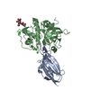
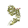
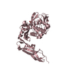
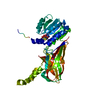
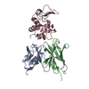
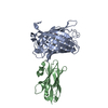
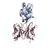
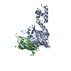
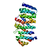
| Title | Human P-cadherin MEC1 with scFv TSP7 bound | ||||||
 Components Components |
| ||||||
 Keywords Keywords | CELL ADHESION/IMMUNE SYSTEM / Cadherin Cell adhesion Antibody Protein-protein interaction /  OXIDOREDUCTASE / CELL ADHESION-IMMUNE SYSTEM complex OXIDOREDUCTASE / CELL ADHESION-IMMUNE SYSTEM complex | ||||||
| Function / homology |  Function and homology information Function and homology informationnegative regulation of timing of catagen / positive regulation of melanosome transport / hair cycle process / positive regulation of tyrosinase activity / positive regulation of keratinocyte proliferation / positive regulation of melanin biosynthetic process / cell-cell adhesion mediated by cadherin / calcium-dependent cell-cell adhesion via plasma membrane cell adhesion molecules / Adherens junctions interactions / catenin complex ...negative regulation of timing of catagen / positive regulation of melanosome transport / hair cycle process / positive regulation of tyrosinase activity / positive regulation of keratinocyte proliferation / positive regulation of melanin biosynthetic process / cell-cell adhesion mediated by cadherin / calcium-dependent cell-cell adhesion via plasma membrane cell adhesion molecules / Adherens junctions interactions / catenin complex / retina homeostasis / cell-cell junction assembly / adherens junction organization / positive regulation of insulin-like growth factor receptor signaling pathway / keratinization / homophilic cell adhesion via plasma membrane adhesion molecules / visual perception / adherens junction / negative regulation of transforming growth factor beta receptor signaling pathway / cell morphogenesis / positive regulation of canonical Wnt signaling pathway / cell junction / cell adhesion / response to xenobiotic stimulus / cadherin binding / calcium ion binding / positive regulation of gene expression / plasma membrane / cytoplasmSimilarity search - Function | ||||||
| Biological species |  Homo sapiens (human) Homo sapiens (human) Mus musculus (house mouse) Mus musculus (house mouse) | ||||||
| Method | X-RAY DIFFRACTION / SYNCHROTRON / MOLECULAR REPLACEMENT / Resolution: 2.55 Å | ||||||
 Authors Authors | Caaveiro, J.M.M. / Kudo, S. / Tsumoto, K. | ||||||
 Citation Citation | Journal: Sci Rep / Year: 2017 Title: Disruption of cell adhesion by an antibody targeting the cell-adhesive intermediate (X-dimer) of human P-cadherin Authors: Kudo, S. / Caaveiro, J.M.M. / Nagatoishi, S. / Miyafusa, T. / Matsuura, T. / Sudou, Y. / Tsumoto, K. | ||||||
| History |
|
- Structure visualization
| Structure viewer | Molecule: MolmilJmol/JSmol |
|---|
- Downloads & links
-Download
| PDBx/mmCIF format | 5jyl.cif.gz | 263.6 KB | Display | PDBx/mmCIF format |
|---|---|---|---|---|
| PDB format | pdb5jyl.ent.gz | 212 KB | Display | PDB format |
| PDBx/mmJSON format | 5jyl.json.gz | Tree view | PDBx/mmJSON format | |
| Others |  Other downloads Other downloads |
-Validation report
| Arichive directory | https://data.pdbj.org/pub/pdb/validation_reports/jy/5jylftp://data.pdbj.org/pub/pdb/validation_reports/jy/5jyl | HTTPS FTP |
|---|
-Related structure data
| Related structure data |  5jymC  1qokS C: citing same article ( S: Starting model for refinement |
|---|---|
| Similar structure data |

-Links
 PDBj
PDBj


- Assembly
| Deposited unit | 
| ||||||||||||||||||||||||||||||||||||||||||||||||||||||||||||||||||||
|---|---|---|---|---|---|---|---|---|---|---|---|---|---|---|---|---|---|---|---|---|---|---|---|---|---|---|---|---|---|---|---|---|---|---|---|---|---|---|---|---|---|---|---|---|---|---|---|---|---|---|---|---|---|---|---|---|---|---|---|---|---|---|---|---|---|---|---|---|---|
| 1 | 
| ||||||||||||||||||||||||||||||||||||||||||||||||||||||||||||||||||||
| 2 | 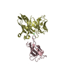
| ||||||||||||||||||||||||||||||||||||||||||||||||||||||||||||||||||||
| Unit cell |
| ||||||||||||||||||||||||||||||||||||||||||||||||||||||||||||||||||||
| Noncrystallographic symmetry (NCS) | NCS domain:
NCS domain segments: Component-ID: 0 / Refine code: 0
NCS ensembles :
|
-Components
-Protein / Antibody , 2 types, 4 molecules ACBD
| #1: Protein | / Placental cadherin / P-cadherin Mass: 11138.497 Da / Num. of mol.: 2 / Fragment: UNP residues 108-207 Source method: isolated from a genetically manipulated source Source: (gene. exp.) Homo sapiens (human) / Gene: CDH3 / Plasmid: pET28 / Production host:  Escherichia coli BL21(DE3) (bacteria) / Strain (production host): BL21 (DE3) / Variant (production host): Rosetta2 / References: UniProt: P22223 Escherichia coli BL21(DE3) (bacteria) / Strain (production host): BL21 (DE3) / Variant (production host): Rosetta2 / References: UniProt: P22223#2: Antibody | Mass: 27346.043 Da / Num. of mol.: 2 Source method: isolated from a genetically manipulated source Source: (gene. exp.) Mus musculus (house mouse) / Plasmid: pRA2 / Production host: Escherichia coli BL21(DE3) (bacteria) / Strain (production host): BL21 (DE3) |
|---|
-Non-polymers , 4 types, 88 molecules 


| #3: Chemical | ChemComp-GOL / Glycerol Mass: 92.094 Da / Num. of mol.: 1 / Source method: obtained synthetically / Formula: C3H8O3 Mass: 92.094 Da / Num. of mol.: 1 / Source method: obtained synthetically / Formula: C3H8O3 | ||
|---|---|---|---|
| #4: Chemical | ChemComp-PO4 / Phosphate Mass: 94.971 Da / Num. of mol.: 1 / Source method: obtained synthetically / Formula: PO4 Mass: 94.971 Da / Num. of mol.: 1 / Source method: obtained synthetically / Formula: PO4 | ||
| #5: Chemical | Chloride Mass: 35.453 Da / Num. of mol.: 2 / Source method: obtained synthetically / Formula: Cl Mass: 35.453 Da / Num. of mol.: 2 / Source method: obtained synthetically / Formula: Cl#6: Water | ChemComp-HOH / | WaterMass: 18.015 Da / Num. of mol.: 84 / Source method: isolated from a natural source / Formula: H2O |
-Details
| Sequence details | MET 0 in chain A, C is N-terminal extension to inhibit strand-swap dimerization. About scFv TSP7 ...MET 0 in chain A, C is N-terminal extension to inhibit strand-swap dimerization. About scFv TSP7 (chain B, D), residues 120-138(SAGGGGSGGG |
|---|
-Experimental details
-Experiment
| Experiment | Method: X-RAY DIFFRACTION / Number of used crystals: 1 |
|---|
- Sample preparation
| Crystal | Density Matthews: 2.47 Å3/Da / Density % sol: 50.3 % |
|---|---|
| Crystal grow | Temperature: 293 K / Method: vapor diffusion, hanging drop / Details: 200 mM potassium phosphate monobasic, 20% PEG 3350 |
-Data collection
| Diffraction | Mean temperature: 100 K |
|---|---|
| Diffraction source | Source: SYNCHROTRON / Site: Photon Factory  / Beamline: BL-5A / Wavelength: 1 Å / Beamline: BL-5A / Wavelength: 1 Å |
| Detector | Type: ADSC QUANTUM 315 / Detector: CCD / Date: Jun 19, 2015 |
| Radiation | Protocol: SINGLE WAVELENGTH / Monochromatic (M) / Laue (L): M / Scattering type: x-ray |
| Radiation wavelength | Wavelength: 1 Å / Relative weight: 1 |
| Reflection | Resolution: 2.55→109.26 Å / Num. obs: 23310 / % possible obs: 99.1 % / Redundancy: 5.4 % / CC1/2: 0.994 / Rmerge(I) obs: 0.125 / Net I/σ(I): 10.4 |
| Reflection shell | Resolution: 2.55→2.69 Å / Redundancy: 5.2 % / Rmerge(I) obs: 0.67 / Mean I/σ(I) obs: 2.4 / % possible all: 96.3 |
- Processing
| Software |
| ||||||||||||||||||||||||||||||||||||||||||||||||||||||||||||||||||||||||||||||||||||||||||||||||||||||||||||||||||||||||||||||||||||||||||||||||||||||||||||||||||||||||||||||||||||||
|---|---|---|---|---|---|---|---|---|---|---|---|---|---|---|---|---|---|---|---|---|---|---|---|---|---|---|---|---|---|---|---|---|---|---|---|---|---|---|---|---|---|---|---|---|---|---|---|---|---|---|---|---|---|---|---|---|---|---|---|---|---|---|---|---|---|---|---|---|---|---|---|---|---|---|---|---|---|---|---|---|---|---|---|---|---|---|---|---|---|---|---|---|---|---|---|---|---|---|---|---|---|---|---|---|---|---|---|---|---|---|---|---|---|---|---|---|---|---|---|---|---|---|---|---|---|---|---|---|---|---|---|---|---|---|---|---|---|---|---|---|---|---|---|---|---|---|---|---|---|---|---|---|---|---|---|---|---|---|---|---|---|---|---|---|---|---|---|---|---|---|---|---|---|---|---|---|---|---|---|---|---|---|---|
| Refinement | Method to determine structure: MOLECULAR REPLACEMENT Starting model: 1QOK Resolution: 2.55→109.26 Å / Cor.coef. Fo:Fc: 0.93 / Cor.coef. Fo:Fc free: 0.884 / SU B: 27.018 / SU ML: 0.273 / Cross valid method: THROUGHOUT / ESU R: 0.973 / ESU R Free: 0.339 / Details: HYDROGENS HAVE BEEN ADDED IN THE RIDING POSITIONS
| ||||||||||||||||||||||||||||||||||||||||||||||||||||||||||||||||||||||||||||||||||||||||||||||||||||||||||||||||||||||||||||||||||||||||||||||||||||||||||||||||||||||||||||||||||||||
| Solvent computation | Ion probe radii: 0.8 Å / Shrinkage radii: 0.8 Å / VDW probe radii: 1.2 Å | ||||||||||||||||||||||||||||||||||||||||||||||||||||||||||||||||||||||||||||||||||||||||||||||||||||||||||||||||||||||||||||||||||||||||||||||||||||||||||||||||||||||||||||||||||||||
| Displacement parameters | Biso mean: 43.579 Å2
| ||||||||||||||||||||||||||||||||||||||||||||||||||||||||||||||||||||||||||||||||||||||||||||||||||||||||||||||||||||||||||||||||||||||||||||||||||||||||||||||||||||||||||||||||||||||
| Refinement step | Cycle: LAST / Resolution: 2.55→109.26 Å
| ||||||||||||||||||||||||||||||||||||||||||||||||||||||||||||||||||||||||||||||||||||||||||||||||||||||||||||||||||||||||||||||||||||||||||||||||||||||||||||||||||||||||||||||||||||||
| Refine LS restraints |
|