 Movie
Movie Controller


+ Open data

- Basic information
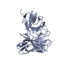
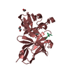
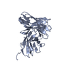
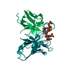
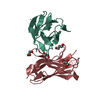
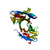
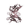
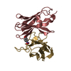
| Entry | Database: PDB / ID: 1qok | ||||||
|---|---|---|---|---|---|---|---|
| Title | MFE-23 AN ANTI-CARCINOEMBRYONIC ANTIGEN SINGLE-CHAIN FV ANTIBODY | ||||||
 Components Components | MFE-23 RECOMBINANT ANTIBODY FRAGMENT | ||||||
 Keywords Keywords |  IMMUNOGLOBULIN / SINGLE-CHAIN FV / ANTI-CARCINOEMBRYONIC ANTIGEN IMMUNOGLOBULIN / SINGLE-CHAIN FV / ANTI-CARCINOEMBRYONIC ANTIGEN | ||||||
| Function / homology |  Function and homology information Function and homology informationImmunoglobulin V-Type / Immunoglobulin V-set domain / Immunoglobulin V-set domain / Immunoglobulin subtype / Immunoglobulin / Ig-like domain profile. / Immunoglobulin-like domain / Immunoglobulin-like domain superfamily / Immunoglobulins / Immunoglobulin-like fold ...Immunoglobulin V-Type / Immunoglobulin V-set domain / Immunoglobulin V-set domain / Immunoglobulin subtype / Immunoglobulin / Ig-like domain profile. / Immunoglobulin-like domain / Immunoglobulin-like domain superfamily / Immunoglobulins / Immunoglobulin-like fold / Immunoglobulin-like / Sandwich / Mainly BetaSimilarity search - Domain/homology | ||||||
| Biological species |  MUS MUSCULUS (house mouse) MUS MUSCULUS (house mouse) | ||||||
| Method | X-RAY DIFFRACTION / MOLECULAR REPLACEMENT / Resolution: 2.4 Å | ||||||
 Authors Authors | Boehm, M.K. / Corper, A.L. / Wan, T. / Sohi, M.K. / Sutton, B.J. / Thornton, J.D. / Keep, P.A. / Chester, K.A. / Begent, R.H.J. / Perkins, S.J. | ||||||
 Citation Citation | Journal: Biochem.J. / Year: 2000 Title: Crystal Structure of the Anti-Carcinoembryonic Antigen Single-Chain Fv Antibody Mfe-23 and a Model for Antigen Binding Based on Intermolecular Contacts Authors: Boehm, M.K. / Corper, A.L. / Wan, T. / Sohi, M.K. / Sutton, B.J. / Thornton, J.D. / Keep, P.A. / Chester, K.A. / Begent, R.H.J. / Perkins, S.J. | ||||||
| History |
|
- Structure visualization
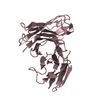
| Structure viewer | Molecule: MolmilJmol/JSmol |
|---|
- Downloads & links
-Download
| PDBx/mmCIF format | 1qok.cif.gz | 60.5 KB | Display | PDBx/mmCIF format |
|---|---|---|---|---|
| PDB format | pdb1qok.ent.gz | 42.5 KB | Display | PDB format |
| PDBx/mmJSON format | 1qok.json.gz | Tree view | PDBx/mmJSON format | |
| Others |  Other downloads Other downloads |
-Validation report
| Arichive directory | https://data.pdbj.org/pub/pdb/validation_reports/qo/1qokftp://data.pdbj.org/pub/pdb/validation_reports/qo/1qok | HTTPS FTP |
|---|
-Related structure data
| Related structure data |  2fbjS S: Starting model for refinement |
|---|---|
| Similar structure data |

-Links
 PDBj
PDBj


- Assembly
| Deposited unit | 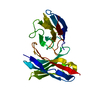
| ||||||||
|---|---|---|---|---|---|---|---|---|---|
| 1 |
| ||||||||
| Unit cell |
|
-Components
| #1: Antibody | Mass: 29874.986 Da / Num. of mol.: 1 / Fragment: SINGLE-CHAIN FV Source method: isolated from a genetically manipulated source Details: ANTI-CARCINOEMBRYONIC ANTIGEN SINGLE-CHAIN FRAGMENT CONTAINING AN N-TERMINAL VH DOMAIN LINKED BY A 3 (4GLY-SER) LINKER TO A VL DOMAIN AND A C-TERMINAL MYC TAG Source: (gene. exp.) MUS MUSCULUS (house mouse) / Plasmid: PUC119 / Cellular location (production host): EXTRACELLULAR / Production host:  ESCHERICHIA COLI (E. coli) / References: GenBank: 2299568, UniProt: Q8K1F2*PLUS ESCHERICHIA COLI (E. coli) / References: GenBank: 2299568, UniProt: Q8K1F2*PLUS |
|---|---|
| #2: Water | ChemComp-HOH / Water Mass: 18.015 Da / Num. of mol.: 96 / Source method: isolated from a natural source / Formula: H2O Mass: 18.015 Da / Num. of mol.: 96 / Source method: isolated from a natural source / Formula: H2O |
| Compound details | SUBDIVISION OF CHAIN: A (C=CHAIN IDENTIFIER; RES1=START RESIDUE; RES1=END RESIDUE;A=ALT CHAIN ...SUBDIVISIO |
| Sequence details | MFE-23 IS A RECOMBINANT PROTEIN THAT WAS PRODUCED FROM THE RANDOM COMBINATION OF A MOUSE VH AND VL ...MFE-23 IS A RECOMBINAN |
-Experimental details
-Experiment
| Experiment | Method: X-RAY DIFFRACTION / Number of used crystals: 1 |
|---|
- Sample preparation
| Crystal | Density Matthews: 2.24 Å3/Da / Density % sol: 43 % | ||||||||||||||||||||||||||||||
|---|---|---|---|---|---|---|---|---|---|---|---|---|---|---|---|---|---|---|---|---|---|---|---|---|---|---|---|---|---|---|---|
| Crystal grow | Temperature: 291 K / Method: vapor diffusion, hanging drop / pH: 6.5 Details: CRYSTALS WERE GROWN BY THE HANGING-DROP VAPOUR DIFFUSION METHOD AT 18 DEGREES (CELSIUS). PROTEIN SOLUTION (2 MG/ML) WAS MIXED 1:1 WITH 100 MM TRIS-HCL (PH6.5) CONTAINING 45% SATURATED ...Details: CRYSTALS WERE GROWN BY THE HANGING-DROP VAPOUR DIFFUSION METHOD AT 18 DEGREES (CELSIUS). PROTEIN SOLUTION (2 MG/ML) WAS MIXED 1:1 WITH 100 MM TRIS-HCL (PH6.5) CONTAINING 45% SATURATED AMMONIUM SULPHATE. A 10 UL DROPLET OF THIS MIXTURE WAS EQULIBRATED AGAINST 0.5 ML 100 MM TRIS-HCL (PH6.5) IN 45% AMMONIUM SULPHATE., pH 6.50 | ||||||||||||||||||||||||||||||
| Crystal grow | *PLUS Temperature: 18 ℃ / Method: vapor diffusion, hanging drop | ||||||||||||||||||||||||||||||
| Components of the solutions | *PLUS
|
-Data collection
| Diffraction | Mean temperature: 291 K |
|---|---|
| Diffraction source | Source: ROTATING ANODE / Type: RIGAKU RUH2R / Wavelength: 1.5418 |
| Detector | Type: R-AXIS II / Detector: IMAGE PLATE / Date: Mar 15, 1994 / Details: COLLIMATOR |
| Radiation | Monochromator: GRAPHITE(002) / Protocol: SINGLE WAVELENGTH / Monochromatic (M) / Laue (L): M / Scattering type: x-ray |
| Radiation wavelength | Wavelength: 1.5418 Å / Relative weight: 1 |
| Reflection | Resolution: 2.4→15 Å / Num. obs: 11539 / % possible obs: 99.6 % / Observed criterion σ(I): 3 / Redundancy: 5.3 % / Biso Wilson estimate: 30.7 Å2 / Rmerge(I) obs: 0.082 / Net I/σ(I): 4.4 |
| Reflection shell | Resolution: 2.4→2.53 Å / Redundancy: 5.1 % / Rmerge(I) obs: 0.189 / Mean I/σ(I) obs: 3.2 / % possible all: 100 |
| Reflection shell | *PLUS % possible obs: 100 % / Num. unique obs: 1613 |
- Processing
| Software |
| ||||||||||||||||||||||||||||||||||||||||||||||||||||||||||||
|---|---|---|---|---|---|---|---|---|---|---|---|---|---|---|---|---|---|---|---|---|---|---|---|---|---|---|---|---|---|---|---|---|---|---|---|---|---|---|---|---|---|---|---|---|---|---|---|---|---|---|---|---|---|---|---|---|---|---|---|---|---|
| Refinement | Method to determine structure: MOLECULAR REPLACEMENT Starting model: 2FBJ Resolution: 2.4→8 Å / Rfactor Rfree error: 0.009 Cross valid method: THROUGHOUT EXCEPT FOR FINAL REFINEMENT CYCLE σ(F): 2 Details: IN THE FINAL REFINEMENT CYCLE, THE WORKING AND TEST SETS WERE MERGED. AFTER THIS REFINEMENT, THE MODEL HAD AN R-FACTOR OF 19.0% AGAINST ALL REFLECTIONS BETWEEN 8.0 AND 2.4 ANGSTROMS..
| ||||||||||||||||||||||||||||||||||||||||||||||||||||||||||||
| Displacement parameters | Biso mean: 25.1 Å2 | ||||||||||||||||||||||||||||||||||||||||||||||||||||||||||||
| Refinement step | Cycle: LAST / Resolution: 2.4→8 Å
| ||||||||||||||||||||||||||||||||||||||||||||||||||||||||||||
| Refine LS restraints |
| ||||||||||||||||||||||||||||||||||||||||||||||||||||||||||||
| LS refinement shell | Resolution: 2.4→2.48 Å / Rfactor Rfree error: 0.043 / Total num. of bins used: 10
| ||||||||||||||||||||||||||||||||||||||||||||||||||||||||||||
| Xplor file |
| ||||||||||||||||||||||||||||||||||||||||||||||||||||||||||||
| Software | *PLUS Name: X-PLOR / Version: 3.1 / Classification: refinement | ||||||||||||||||||||||||||||||||||||||||||||||||||||||||||||
| Refinement | *PLUS Num. reflection obs: 11204 / Num. reflection Rfree: 909 / % reflection Rfree: 8.5 % / Rfactor all: 0.19 | ||||||||||||||||||||||||||||||||||||||||||||||||||||||||||||
| Solvent computation | *PLUS | ||||||||||||||||||||||||||||||||||||||||||||||||||||||||||||
| Displacement parameters | *PLUS | ||||||||||||||||||||||||||||||||||||||||||||||||||||||||||||
| Refine LS restraints | *PLUS
| ||||||||||||||||||||||||||||||||||||||||||||||||||||||||||||
| LS refinement shell | *PLUS Rfactor Rfree: 0.4 |
