 Movie
Movie Controller


+ Open data

- Basic information
| Entry | Database: PDB / ID: 5jxe | ||||||
|---|---|---|---|---|---|---|---|
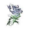
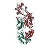
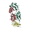
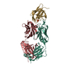
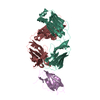
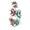
| Title | Human PD-1 ectodomain complexed with Pembrolizumab Fab | ||||||
 Components Components |
| ||||||
 Keywords Keywords |  IMMUNE SYSTEM / human PD-1 Pembrolizumab Fab IMMUNE SYSTEM / human PD-1 Pembrolizumab Fab | ||||||
| Function / homology |  Function and homology information Function and homology informationnegative regulation of tolerance induction / regulatory T cell apoptotic process / negative regulation of immune response / B cell apoptotic process / positive regulation of T cell apoptotic process / negative regulation of B cell apoptotic process / humoral immune response / PD-1 signaling / regulation of immune response / Potential therapeutics for SARS ...negative regulation of tolerance induction / regulatory T cell apoptotic process / negative regulation of immune response / B cell apoptotic process / positive regulation of T cell apoptotic process / negative regulation of B cell apoptotic process / humoral immune response / PD-1 signaling / regulation of immune response / Potential therapeutics for SARS / adaptive immune response / external side of plasma membrane / apoptotic process / plasma membraneSimilarity search - Function | ||||||
| Biological species |  Homo sapiens (human) Homo sapiens (human) | ||||||
| Method | X-RAY DIFFRACTION / SYNCHROTRON / MOLECULAR REPLACEMENT / Resolution: 2.9 Å | ||||||
 Authors Authors | Na, Z. / Bharath, S.R. / Song, H. | ||||||
 Citation Citation | Journal: Cell Res. / Year: 2017 Title: Structural basis for blocking PD-1-mediated immune suppression by therapeutic antibody pembrolizumab. Authors: Na, Z. / Yeo, S.P. / Bharath, S.R. / Bowler, M.W. / Balijkcij, E. / Wang, C.I. / Song, H. | ||||||
| History |
|
- Structure visualization
| Structure viewer | Molecule: MolmilJmol/JSmol |
|---|
- Downloads & links
-Download
| PDBx/mmCIF format | 5jxe.cif.gz | 208.4 KB | Display | PDBx/mmCIF format |
|---|---|---|---|---|
| PDB format | pdb5jxe.ent.gz | 164.3 KB | Display | PDB format |
| PDBx/mmJSON format | 5jxe.json.gz | Tree view | PDBx/mmJSON format | |
| Others |  Other downloads Other downloads |
-Validation report
| Arichive directory | https://data.pdbj.org/pub/pdb/validation_reports/jx/5jxeftp://data.pdbj.org/pub/pdb/validation_reports/jx/5jxe | HTTPS FTP |
|---|
-Related structure data


-Links
 PDBj
PDBj


- Assembly
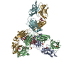
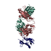
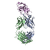
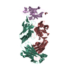
| Deposited unit | 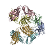
| ||||||||
|---|---|---|---|---|---|---|---|---|---|
| 1 | 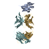
| ||||||||
| 2 | 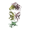
| ||||||||
| Unit cell |
|
-Components
| #1: Protein | / hPD-1 Mass: 12714.137 Da / Num. of mol.: 2 / Fragment: UNP RESIDUES 34-146 Source method: isolated from a genetically manipulated source Source: (gene. exp.) Homo sapiens (human) / Gene: PDCD1, PD1 / Production host:  Escherichia coli (E. coli) / References: UniProt: Q15116 Escherichia coli (E. coli) / References: UniProt: Q15116#2: Antibody | Mass: 23768.449 Da / Num. of mol.: 2 Source method: isolated from a genetically manipulated source Source: (gene. exp.) Homo sapiens (human) / Production host: Homo sapiens (human)#3: Antibody | Mass: 23681.471 Da / Num. of mol.: 2 Source method: isolated from a genetically manipulated source Source: (gene. exp.) Homo sapiens (human) / Production host: Homo sapiens (human)#4: Water | ChemComp-HOH / | Water Mass: 18.015 Da / Num. of mol.: 10 / Source method: isolated from a natural source / Formula: H2O Mass: 18.015 Da / Num. of mol.: 10 / Source method: isolated from a natural source / Formula: H2O |
|---|
-Experimental details
-Experiment
| Experiment | Method: X-RAY DIFFRACTION / Number of used crystals: 1 |
|---|
- Sample preparation
| Crystal | Density Matthews: 2.54 Å3/Da / Density % sol: 51.61 % |
|---|---|
| Crystal grow | Temperature: 277 K / Method: vapor diffusion, hanging drop Details: 0.1 M sodium citrate pH 5.6, 19 mM n-Decyl-N,N-dimethylglycine, 20% isopropanol and 20% PEG 4000 |
-Data collection
| Diffraction | Mean temperature: 100 K |
|---|---|
| Diffraction source | Source: SYNCHROTRON / Site: ESRF  / Beamline: MASSIF-1 / Wavelength: 0.966 Å / Beamline: MASSIF-1 / Wavelength: 0.966 Å |
| Detector | Type: DECTRIS PILATUS 2M / Detector: PIXEL / Date: Jan 27, 2016 / Details: Si with Pt coating |
| Radiation | Protocol: SINGLE WAVELENGTH / Monochromatic (M) / Laue (L): M / Scattering type: x-ray |
| Radiation wavelength | Wavelength: 0.966 Å / Relative weight: 1 |
| Reflection | Resolution: 2.9→29.926 Å / Num. obs: 28657 / % possible obs: 99.4 % / Redundancy: 4.3 % / Biso Wilson estimate: 67.2 Å2 / CC1/2: 0.995 / Rmerge(I) obs: 0.12 / Net I/σ(I): 9 |
| Reflection shell | Resolution: 2.9→3.08 Å / Redundancy: 4.2 % / Mean I/σ(I) obs: 1.5 / % possible all: 99.2 |
- Processing
| Software |
| |||||||||||||||||||||||||||||||||||||||||||||||||||||||||||||||||||||||||||||
|---|---|---|---|---|---|---|---|---|---|---|---|---|---|---|---|---|---|---|---|---|---|---|---|---|---|---|---|---|---|---|---|---|---|---|---|---|---|---|---|---|---|---|---|---|---|---|---|---|---|---|---|---|---|---|---|---|---|---|---|---|---|---|---|---|---|---|---|---|---|---|---|---|---|---|---|---|---|---|
| Refinement | Method to determine structure: MOLECULAR REPLACEMENT Starting model: 4ZQK, 5DK3 Resolution: 2.9→29.926 Å / SU ML: 0.4 / Cross valid method: FREE R-VALUE / σ(F): 1.34 / Phase error: 30.97
| |||||||||||||||||||||||||||||||||||||||||||||||||||||||||||||||||||||||||||||
| Solvent computation | Shrinkage radii: 0.9 Å / VDW probe radii: 1.11 Å | |||||||||||||||||||||||||||||||||||||||||||||||||||||||||||||||||||||||||||||
| Refinement step | Cycle: LAST / Resolution: 2.9→29.926 Å
| |||||||||||||||||||||||||||||||||||||||||||||||||||||||||||||||||||||||||||||
| Refine LS restraints |
| |||||||||||||||||||||||||||||||||||||||||||||||||||||||||||||||||||||||||||||
| LS refinement shell |
|
