 Movie
Movie Controller


+ Open data

- Basic information
| Entry | Database: PDB / ID: 5jmr | ||||||
|---|---|---|---|---|---|---|---|
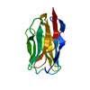
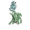
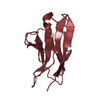
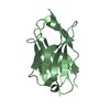
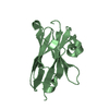
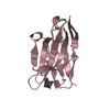
| Title | X-ray structure of the furin inhibitory antibody Nb14 | ||||||
 Components Components | camelid VHH fragment | ||||||
 Keywords Keywords |  IMMUNE SYSTEM / camelid / antibody / inhibitor / furin IMMUNE SYSTEM / camelid / antibody / inhibitor / furin | ||||||
| Function / homology | Immunoglobulins / Immunoglobulin-like / Sandwich / Mainly Beta Function and homology information Function and homology information | ||||||
| Biological species |  Camelus dromedarius (Arabian camel) Camelus dromedarius (Arabian camel) | ||||||
| Method | X-RAY DIFFRACTION / SYNCHROTRON / MOLECULAR REPLACEMENT / Resolution: 2.27 Å | ||||||
 Authors Authors | Dahms, S.O. / Than, M.E. | ||||||
 Citation Citation | Journal: Sci Rep / Year: 2016 Title: The structure of a furin-antibody complex explains non-competitive inhibition by steric exclusion of substrate conformers. Authors: Dahms, S.O. / Creemers, J.W. / Schaub, Y. / Bourenkov, G.P. / Zogg, T. / Brandstetter, H. / Than, M.E. | ||||||
| History |
|
- Structure visualization
| Structure viewer | Molecule: MolmilJmol/JSmol |
|---|
- Downloads & links
-Download
| PDBx/mmCIF format | 5jmr.cif.gz | 60.2 KB | Display | PDBx/mmCIF format |
|---|---|---|---|---|
| PDB format | pdb5jmr.ent.gz | 43.3 KB | Display | PDB format |
| PDBx/mmJSON format | 5jmr.json.gz | Tree view | PDBx/mmJSON format | |
| Others |  Other downloads Other downloads |
-Validation report
| Arichive directory | https://data.pdbj.org/pub/pdb/validation_reports/jm/5jmrftp://data.pdbj.org/pub/pdb/validation_reports/jm/5jmr | HTTPS FTP |
|---|
-Related structure data
| Related structure data |  5jmoC 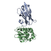 1op9S S: Starting model for refinement C: citing same article ( |
|---|---|
| Similar structure data |


-Links
 PDBj
PDBj

- Assembly
| Deposited unit | 
| ||||||||
|---|---|---|---|---|---|---|---|---|---|
| 1 | 
| ||||||||
| 2 | 
| ||||||||
| Unit cell |
| ||||||||
| Components on special symmetry positions |
|
-Components
| #1: Antibody | Mass: 13646.156 Da / Num. of mol.: 2 Source method: isolated from a genetically manipulated source Source: (gene. exp.) Camelus dromedarius (Arabian camel) / Production host:  Escherichia coli (E. coli) Escherichia coli (E. coli)#2: Chemical | Chloride  Mass: 35.453 Da / Num. of mol.: 2 / Source method: obtained synthetically / Formula: Cl Mass: 35.453 Da / Num. of mol.: 2 / Source method: obtained synthetically / Formula: Cl#3: Water | ChemComp-HOH / | Water Mass: 18.015 Da / Num. of mol.: 131 / Source method: isolated from a natural source / Formula: H2O Mass: 18.015 Da / Num. of mol.: 131 / Source method: isolated from a natural source / Formula: H2O |
|---|
-Experimental details
-Experiment
| Experiment | Method: X-RAY DIFFRACTION / Number of used crystals: 1 |
|---|
- Sample preparation
| Crystal | Density Matthews: 4.04 Å3/Da / Density % sol: 69.58 % |
|---|---|
| Crystal grow | Temperature: 293.15 K / Method: evaporation / Details: 10 mM Hepes, pH 7.5, 100 mM NaCl and 1mM CaCl2 |
-Data collection
| Diffraction | Mean temperature: 100 K |
|---|---|
| Diffraction source | Source: SYNCHROTRON / Site: BESSY  / Beamline: 14.1 / Wavelength: 0.918409 Å / Beamline: 14.1 / Wavelength: 0.918409 Å |
| Detector | Type: DECTRIS PILATUS 6M / Detector: PIXEL / Date: Jun 16, 2014 |
| Radiation | Protocol: SINGLE WAVELENGTH / Monochromatic (M) / Laue (L): M / Scattering type: x-ray |
| Radiation wavelength | Wavelength: 0.918409 Å / Relative weight: 1 |
| Reflection | Resolution: 2.27→35.356 Å / Num. obs: 21253 / % possible obs: 99.7 % / Redundancy: 6.6 % / Net I/σ(I): 11.9 |
- Processing
| Software |
| |||||||||||||||||||||||||||||||||||||||||||||||||||||||||||||||
|---|---|---|---|---|---|---|---|---|---|---|---|---|---|---|---|---|---|---|---|---|---|---|---|---|---|---|---|---|---|---|---|---|---|---|---|---|---|---|---|---|---|---|---|---|---|---|---|---|---|---|---|---|---|---|---|---|---|---|---|---|---|---|---|---|
| Refinement | Method to determine structure: MOLECULAR REPLACEMENT Starting model: 1OP9 Resolution: 2.27→35.356 Å / SU ML: 0.23 / Cross valid method: FREE R-VALUE / σ(F): 1.35 / Phase error: 21.96 / Stereochemistry target values: ML
| |||||||||||||||||||||||||||||||||||||||||||||||||||||||||||||||
| Solvent computation | Shrinkage radii: 0.9 Å / VDW probe radii: 1.11 Å / Solvent model: FLAT BULK SOLVENT MODEL | |||||||||||||||||||||||||||||||||||||||||||||||||||||||||||||||
| Refinement step | Cycle: LAST / Resolution: 2.27→35.356 Å
| |||||||||||||||||||||||||||||||||||||||||||||||||||||||||||||||
| Refine LS restraints |
| |||||||||||||||||||||||||||||||||||||||||||||||||||||||||||||||
| LS refinement shell |
|
