 Movie
Movie Controller

[English] 日本語
 Yorodumi
- PDB-5ixc: Human GIVD cytosolic phospholipase A2 in complex with Methyl gamm... -
+ Open data

- Basic information
| Entry | Database: PDB / ID: 5ixc | ||||||
|---|---|---|---|---|---|---|---|
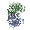
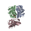
| Title | Human GIVD cytosolic phospholipase A2 in complex with Methyl gamma-Linolenyl Fluorophosphonate | ||||||
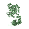
 Components Components | Cytosolic phospholipase A2 delta | ||||||
 Keywords Keywords | HYDROLASE/HYDROLASE inhibitor / Inhibitor /  Signal Transduction / Phospholipase / alpha/beta hydrolase / Calcium binding / C2 domain / HYDROLASE / HYDROLASE-HYDROLASE inhibitor complex Signal Transduction / Phospholipase / alpha/beta hydrolase / Calcium binding / C2 domain / HYDROLASE / HYDROLASE-HYDROLASE inhibitor complex | ||||||
| Function / homology |  Function and homology information Function and homology informationphosphatidylglycerol acyl-chain remodeling / phosphatidylinositol acyl-chain remodeling / glycerophospholipid catabolic process / Hydrolysis of LPC / Acyl chain remodelling of PG / phospholipase A1 activity / Acyl chain remodelling of PC / Acyl chain remodelling of PI / Acyl chain remodelling of PS / Acyl chain remodelling of PE ...phosphatidylglycerol acyl-chain remodeling / phosphatidylinositol acyl-chain remodeling / glycerophospholipid catabolic process / Hydrolysis of LPC / Acyl chain remodelling of PG / phospholipase A1 activity / Acyl chain remodelling of PC / Acyl chain remodelling of PI / Acyl chain remodelling of PS / Acyl chain remodelling of PE / Synthesis of PA / calcium-dependent phospholipid binding / calcium-dependent phospholipase A2 activity / phospholipase A2 / fatty acid metabolic process / calcium ion binding / membrane / cytosolSimilarity search - Function | ||||||
| Biological species |  Homo sapiens (human) Homo sapiens (human) | ||||||
| Method | X-RAY DIFFRACTION / SYNCHROTRON / MOLECULAR REPLACEMENT / Resolution: 2.65 Å | ||||||
 Authors Authors | Wang, H. / Klein, M.G. | ||||||
 Citation Citation | Journal: J.Mol.Biol. / Year: 2016 Title: Structure of Human GIVD Cytosolic Phospholipase A2 Reveals Insights into Substrate Recognition. Authors: Wang, H. / Klein, M.G. / Snell, G. / Lane, W. / Zou, H. / Levin, I. / Li, K. / Sang, B.C. | ||||||
| History |
|
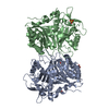
- Structure visualization
| Structure viewer | Molecule: MolmilJmol/JSmol |
|---|
- Downloads & links
-Download
| PDBx/mmCIF format | 5ixc.cif.gz | 286.4 KB | Display | PDBx/mmCIF format |
|---|---|---|---|---|
| PDB format | pdb5ixc.ent.gz | 228.2 KB | Display | PDB format |
| PDBx/mmJSON format | 5ixc.json.gz | Tree view | PDBx/mmJSON format | |
| Others |  Other downloads Other downloads |
-Validation report
| Arichive directory | https://data.pdbj.org/pub/pdb/validation_reports/ix/5ixcftp://data.pdbj.org/pub/pdb/validation_reports/ix/5ixc | HTTPS FTP |
|---|
-Related structure data
| Related structure data |  5iz5C  5izrSC C: citing same article ( S: Starting model for refinement |
|---|---|
| Similar structure data |


-Links
 PDBj
PDBj


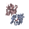
- Assembly
| Deposited unit | 
| ||||||||||||||||||
|---|---|---|---|---|---|---|---|---|---|---|---|---|---|---|---|---|---|---|---|
| 1 | 
| ||||||||||||||||||
| 2 | 
| ||||||||||||||||||
| Unit cell |
| ||||||||||||||||||
| Noncrystallographic symmetry (NCS) | NCS domain:
NCS domain segments: Component-ID: 0 / Ens-ID: 1 / Beg auth comp-ID: GLN / Beg label comp-ID: GLN / End auth comp-ID: THR / End label comp-ID: THR / Refine code: 0 / Auth seq-ID: 15 - 808 / Label seq-ID: 19 - 812
|
-Components
| #1: Protein | Mass: 91444.305 Da / Num. of mol.: 2 / Fragment: UNP residues 2-810 Source method: isolated from a genetically manipulated source Source: (gene. exp.) Homo sapiens (human) / Gene: PLA2G4D / Production host:   Spodoptera frugiperda (fall armyworm) / References: UniProt: Q86XP0, phospholipase A2 Spodoptera frugiperda (fall armyworm) / References: UniProt: Q86XP0, phospholipase A2#2: Chemical |   Mass: 344.444 Da / Num. of mol.: 2 / Source method: obtained synthetically / Formula: C19H34FO2P Mass: 344.444 Da / Num. of mol.: 2 / Source method: obtained synthetically / Formula: C19H34FO2P#3: Chemical | ChemComp-BA /   Mass: 137.327 Da / Num. of mol.: 6 / Source method: obtained synthetically / Formula: Ba Mass: 137.327 Da / Num. of mol.: 6 / Source method: obtained synthetically / Formula: Ba#4: Water | ChemComp-HOH / | Water Mass: 18.015 Da / Num. of mol.: 71 / Source method: isolated from a natural source / Formula: H2O Mass: 18.015 Da / Num. of mol.: 71 / Source method: isolated from a natural source / Formula: H2O |
|---|
-Experimental details
-Experiment
| Experiment | Method: X-RAY DIFFRACTION / Number of used crystals: 1 |
|---|
- Sample preparation
| Crystal | Density Matthews: 2.4 Å3/Da / Density % sol: 48.7 % |
|---|---|
| Crystal grow | Temperature: 277 K / Method: vapor diffusion, hanging drop Details: 100 mM MES (pH 6.0-6.2), 50 mM sodium acetate, and 10-14% (v/v) PEG20K PH range: 6.0-6.2 |
-Data collection
| Diffraction | Mean temperature: 100 K |
|---|---|
| Diffraction source | Source: SYNCHROTRON / Site: ALS  / Beamline: 5.0.3 / Wavelength: 0.997648 Å / Beamline: 5.0.3 / Wavelength: 0.997648 Å |
| Detector | Type: ADSC QUANTUM 210r / Detector: CCD / Date: Apr 18, 2015 |
| Radiation | Protocol: SINGLE WAVELENGTH / Monochromatic (M) / Laue (L): M / Scattering type: x-ray |
| Radiation wavelength | Wavelength: 0.997648 Å / Relative weight: 1 |
| Reflection | Resolution: 2.65→90.97 Å / Num. obs: 52014 / % possible obs: 100 % / Redundancy: 8.1 % / Rmerge(I) obs: 0.092 / Net I/σ(I): 20 |
| Reflection shell | Resolution: 2.65→2.7 Å / Redundancy: 8.1 % / Mean I/σ(I) obs: 2.1 / % possible all: 100 |
- Processing
| Software |
| ||||||||||||||||||||||||||||||||||||||||||||||||||||||||||||||||||||||||||||||||||||||||||||||||||||||||||||||||||||||||||||||||||||||||||||||||||||||||||||||||||||||||||||||||||||||
|---|---|---|---|---|---|---|---|---|---|---|---|---|---|---|---|---|---|---|---|---|---|---|---|---|---|---|---|---|---|---|---|---|---|---|---|---|---|---|---|---|---|---|---|---|---|---|---|---|---|---|---|---|---|---|---|---|---|---|---|---|---|---|---|---|---|---|---|---|---|---|---|---|---|---|---|---|---|---|---|---|---|---|---|---|---|---|---|---|---|---|---|---|---|---|---|---|---|---|---|---|---|---|---|---|---|---|---|---|---|---|---|---|---|---|---|---|---|---|---|---|---|---|---|---|---|---|---|---|---|---|---|---|---|---|---|---|---|---|---|---|---|---|---|---|---|---|---|---|---|---|---|---|---|---|---|---|---|---|---|---|---|---|---|---|---|---|---|---|---|---|---|---|---|---|---|---|---|---|---|---|---|---|---|
| Refinement | Method to determine structure: MOLECULAR REPLACEMENT Starting model: 5IZR Resolution: 2.65→90.97 Å / Cor.coef. Fo:Fc: 0.939 / Cor.coef. Fo:Fc free: 0.928 / SU B: 12.931 / SU ML: 0.26 / Cross valid method: THROUGHOUT / ESU R: 0.905 / ESU R Free: 0.319 / Stereochemistry target values: MAXIMUM LIKELIHOOD / Details: HYDROGENS HAVE BEEN ADDED IN THE RIDING POSITIONS
| ||||||||||||||||||||||||||||||||||||||||||||||||||||||||||||||||||||||||||||||||||||||||||||||||||||||||||||||||||||||||||||||||||||||||||||||||||||||||||||||||||||||||||||||||||||||
| Solvent computation | Ion probe radii: 0.8 Å / Shrinkage radii: 0.8 Å / VDW probe radii: 1.2 Å / Solvent model: MASK | ||||||||||||||||||||||||||||||||||||||||||||||||||||||||||||||||||||||||||||||||||||||||||||||||||||||||||||||||||||||||||||||||||||||||||||||||||||||||||||||||||||||||||||||||||||||
| Displacement parameters | Biso mean: 65.842 Å2
| ||||||||||||||||||||||||||||||||||||||||||||||||||||||||||||||||||||||||||||||||||||||||||||||||||||||||||||||||||||||||||||||||||||||||||||||||||||||||||||||||||||||||||||||||||||||
| Refinement step | Cycle: LAST / Resolution: 2.65→90.97 Å
| ||||||||||||||||||||||||||||||||||||||||||||||||||||||||||||||||||||||||||||||||||||||||||||||||||||||||||||||||||||||||||||||||||||||||||||||||||||||||||||||||||||||||||||||||||||||
| Refine LS restraints |
|