 Movie
Movie Controller

[English] 日本語
 Yorodumi
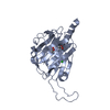
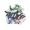
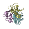
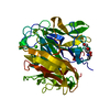
- PDB-5hzf: Single Chain Recombinant Globular Head of the Complement System P... -
+ Open data

- Basic information
| Entry | Database: PDB / ID: 5hzf | |||||||||
|---|---|---|---|---|---|---|---|---|---|---|
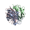
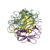
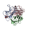
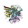
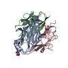
| Title | Single Chain Recombinant Globular Head of the Complement System Protein C1q in complex with magnesium | |||||||||
 Components Components | Complement C1q subcomponent subunit A,Complement C1q subcomponent subunit C,Complement C1q subcomponent subunit B | |||||||||
 Keywords Keywords |  SIGNALING PROTEIN / gC1q domain / complement / C1q SIGNALING PROTEIN / gC1q domain / complement / C1q | |||||||||
| Function / homology |  Function and homology information Function and homology informationcomplement component C1 complex / complement component C1q complex / negative regulation of macrophage differentiation / synapse pruning / negative regulation of granulocyte differentiation / vertebrate eye-specific patterning / complement-mediated synapse pruning / collagen trimer / complement activation / Classical antibody-mediated complement activation ...complement component C1 complex / complement component C1q complex / negative regulation of macrophage differentiation / synapse pruning / negative regulation of granulocyte differentiation / vertebrate eye-specific patterning / complement-mediated synapse pruning / collagen trimer / complement activation / Classical antibody-mediated complement activation / neuron remodeling / Initial triggering of complement / complement activation, classical pathway / Regulation of Complement cascade / astrocyte activation / synapse organization / microglial cell activation / cell-cell signaling / amyloid-beta binding / postsynapse / collagen-containing extracellular matrix / blood microparticle / immune response / innate immune response / synapse / extracellular space / extracellular regionSimilarity search - Function | |||||||||
| Biological species |  Homo sapiens (human) Homo sapiens (human) | |||||||||
| Method | X-RAY DIFFRACTION / SYNCHROTRON / MOLECULAR REPLACEMENT / Resolution: 1.55 Å | |||||||||
 Authors Authors | Moreau, C.P. / Gaboriaud, C.P. | |||||||||
| Funding support |  France, 1items France, 1items
| |||||||||
 Citation Citation | Journal: Front Immunol / Year: 2016 Title: Structural and Functional Characterization of a Single-Chain Form of the Recognition Domain of Complement Protein C1q. Authors: Moreau, C. / Bally, I. / Chouquet, A. / Bottazzi, B. / Ghebrehiwet, B. / Gaboriaud, C. / Thielens, N. | |||||||||
| History |
|
- Structure visualization
| Structure viewer | Molecule: MolmilJmol/JSmol |
|---|
- Downloads & links
-Download
| PDBx/mmCIF format | 5hzf.cif.gz | 182.2 KB | Display | PDBx/mmCIF format |
|---|---|---|---|---|
| PDB format | pdb5hzf.ent.gz | 144.2 KB | Display | PDB format |
| PDBx/mmJSON format | 5hzf.json.gz | Tree view | PDBx/mmJSON format | |
| Others |  Other downloads Other downloads |
-Validation report
| Arichive directory | https://data.pdbj.org/pub/pdb/validation_reports/hz/5hzfftp://data.pdbj.org/pub/pdb/validation_reports/hz/5hzf | HTTPS FTP |
|---|
-Related structure data
| Related structure data |  5hkjC  1pk6S S: Starting model for refinement C: citing same article ( |
|---|---|
| Similar structure data |


-Links
 PDBj
PDBj

- Assembly
| Deposited unit | 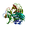
| ||||||||
|---|---|---|---|---|---|---|---|---|---|
| 1 |
| ||||||||
| Unit cell |
|
-Components
| #1: Protein | Mass: 45697.594 Da / Num. of mol.: 1 Source method: isolated from a genetically manipulated source Source: (gene. exp.) Homo sapiens (human) / Gene: C1QA, C1QC, C1QG, C1QB / Plasmid: pcDNA3.1 / Cell line (production host): HEK 293-F / Production host: Homo sapiens (human)References: UniProt: P02745, UniProt: P02747, UniProt: P02746 |
|---|---|
| #2: Chemical | ChemComp-MG /   Mass: 24.305 Da / Num. of mol.: 1 / Source method: obtained synthetically / Formula: Mg Mass: 24.305 Da / Num. of mol.: 1 / Source method: obtained synthetically / Formula: Mg |
| #3: Water | ChemComp-HOH / Water Mass: 18.015 Da / Num. of mol.: 315 / Source method: isolated from a natural source / Formula: H2O Mass: 18.015 Da / Num. of mol.: 315 / Source method: isolated from a natural source / Formula: H2O |
-Experimental details
-Experiment
| Experiment | Method: X-RAY DIFFRACTION / Number of used crystals: 1 |
|---|
- Sample preparation
| Crystal | Density Matthews: 1.94 Å3/Da / Density % sol: 36.79 % |
|---|---|
| Crystal grow | Temperature: 293 K / Method: vapor diffusion, hanging drop / pH: 7.5 / Details: 30% PEG 8000, 0.1M HEPES, pH 7.5 |
-Data collection
| Diffraction | Mean temperature: 100 K |
|---|---|
| Diffraction source | Source: SYNCHROTRON / Site: ESRF / Beamline: ID23-1 / Wavelength: 0.976 Å |
| Detector | Type: DECTRIS PILATUS 6M-F / Detector: PIXEL / Date: Jun 15, 2015 |
| Radiation | Protocol: SINGLE WAVELENGTH / Monochromatic (M) / Laue (L): M / Scattering type: x-ray |
| Radiation wavelength | Wavelength: 0.976 Å / Relative weight: 1 |
| Reflection | Resolution: 1.55→100 Å / Num. obs: 49348 / % possible obs: 98.4 % / Redundancy: 4.9 % / Rsym value: 0.056 / Net I/σ(I): 14.85 |
| Reflection shell | Resolution: 1.55→1.61 Å / Redundancy: 4.7 % / Mean I/σ(I) obs: 1.95 / Rsym value: 0.0753 / % possible all: 96.7 |
- Processing
| Software |
| |||||||||||||||||||||||||||||||||||||||||||||||||||||||||||||||||||||||||||||||||||||||||||||||||||||||||||||||||||||||||||||||||||||
|---|---|---|---|---|---|---|---|---|---|---|---|---|---|---|---|---|---|---|---|---|---|---|---|---|---|---|---|---|---|---|---|---|---|---|---|---|---|---|---|---|---|---|---|---|---|---|---|---|---|---|---|---|---|---|---|---|---|---|---|---|---|---|---|---|---|---|---|---|---|---|---|---|---|---|---|---|---|---|---|---|---|---|---|---|---|---|---|---|---|---|---|---|---|---|---|---|---|---|---|---|---|---|---|---|---|---|---|---|---|---|---|---|---|---|---|---|---|---|---|---|---|---|---|---|---|---|---|---|---|---|---|---|---|---|
| Refinement | Method to determine structure: MOLECULAR REPLACEMENT Starting model: 1PK6 Resolution: 1.55→42.901 Å / SU ML: 0.2 / Cross valid method: FREE R-VALUE / σ(F): 1.34 / Phase error: 21.03
| |||||||||||||||||||||||||||||||||||||||||||||||||||||||||||||||||||||||||||||||||||||||||||||||||||||||||||||||||||||||||||||||||||||
| Solvent computation | Shrinkage radii: 0.9 Å / VDW probe radii: 1.11 Å | |||||||||||||||||||||||||||||||||||||||||||||||||||||||||||||||||||||||||||||||||||||||||||||||||||||||||||||||||||||||||||||||||||||
| Refinement step | Cycle: LAST / Resolution: 1.55→42.901 Å
| |||||||||||||||||||||||||||||||||||||||||||||||||||||||||||||||||||||||||||||||||||||||||||||||||||||||||||||||||||||||||||||||||||||
| Refine LS restraints |
| |||||||||||||||||||||||||||||||||||||||||||||||||||||||||||||||||||||||||||||||||||||||||||||||||||||||||||||||||||||||||||||||||||||
| LS refinement shell |
| |||||||||||||||||||||||||||||||||||||||||||||||||||||||||||||||||||||||||||||||||||||||||||||||||||||||||||||||||||||||||||||||||||||
| Refinement TLS params. | Method: refined / Refine-ID: X-RAY DIFFRACTION
| |||||||||||||||||||||||||||||||||||||||||||||||||||||||||||||||||||||||||||||||||||||||||||||||||||||||||||||||||||||||||||||||||||||
| Refinement TLS group |
|
