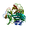
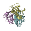
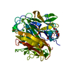
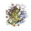
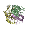
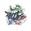
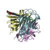
Entry Database : PDB / ID : 2jg8Title Crystallographic structure of human C1q globular heads complexed to phosphatidyl-serine (Complement C1q subcomponent subunit ...) x 3 Keywords / / / / / / / / / / / / / / Function / homology Biological species Homo sapiens (human)Method / / / Resolution : 2.05 Å Authors Paidassi, H. / Tacnet-Delorme, P. / Garlatti, V. / Darnault, C. / Ghebrehiwet, B. / Gaboriaud, C. / Arlaud, G.J. / Frachet, P. Journal : J.Immunol. / Year : 2008Title : C1Q Binds Phosphatidylserine and Likely Acts as a Multiligand-Bridging Molecule in Apoptotic Cell Recognition.Authors : Paidassi, H. / Tacnet-Delorme, P. / Garlatti, V. / Darnault, C. / Ghebrehiwet, B. / Gaboriaud, C. / Arlaud, G.J. / Frachet, P. History Deposition Feb 9, 2007 Deposition site / Processing site Revision 1.0 Feb 19, 2008 Provider / Type Revision 1.1 May 8, 2011 Group Revision 1.2 Jul 13, 2011 Group Revision 1.3 Dec 5, 2018 Group Advisory / Data collection ... Advisory / Data collection / Database references / Source and taxonomy / Structure summary Category citation / entity ... citation / entity / entity_name_com / entity_src_nat / pdbx_entity_src_syn / pdbx_unobs_or_zero_occ_atoms / struct_ref / struct_ref_seq Item _citation.journal_id_ASTM / _citation.journal_id_CSD ... _citation.journal_id_ASTM / _citation.journal_id_CSD / _citation.page_last / _citation.pdbx_database_id_DOI / _entity.pdbx_description / _entity.src_method / _struct_ref.pdbx_align_begin / _struct_ref.pdbx_seq_one_letter_code / _struct_ref_seq.db_align_beg / _struct_ref_seq.db_align_end Revision 1.4 Jul 29, 2020 Group Data collection / Derived calculations ... Data collection / Derived calculations / Other / Structure summary Category chem_comp / entity ... chem_comp / entity / pdbx_chem_comp_identifier / pdbx_database_status / pdbx_entity_nonpoly / pdbx_struct_conn_angle / struct_conn / struct_site / struct_site_gen Item _chem_comp.name / _chem_comp.type ... _chem_comp.name / _chem_comp.type / _entity.pdbx_description / _pdbx_database_status.status_code_sf / _pdbx_entity_nonpoly.name / _pdbx_struct_conn_angle.ptnr1_auth_asym_id / _pdbx_struct_conn_angle.ptnr1_auth_comp_id / _pdbx_struct_conn_angle.ptnr1_auth_seq_id / _pdbx_struct_conn_angle.ptnr1_label_asym_id / _pdbx_struct_conn_angle.ptnr1_label_atom_id / _pdbx_struct_conn_angle.ptnr1_label_comp_id / _pdbx_struct_conn_angle.ptnr1_label_seq_id / _pdbx_struct_conn_angle.ptnr3_auth_asym_id / _pdbx_struct_conn_angle.ptnr3_auth_comp_id / _pdbx_struct_conn_angle.ptnr3_auth_seq_id / _pdbx_struct_conn_angle.ptnr3_label_asym_id / _pdbx_struct_conn_angle.ptnr3_label_atom_id / _pdbx_struct_conn_angle.ptnr3_label_comp_id / _pdbx_struct_conn_angle.ptnr3_label_seq_id / _pdbx_struct_conn_angle.value / _struct_conn.pdbx_dist_value / _struct_conn.ptnr1_auth_asym_id / _struct_conn.ptnr1_auth_comp_id / _struct_conn.ptnr1_auth_seq_id / _struct_conn.ptnr1_label_asym_id / _struct_conn.ptnr1_label_atom_id / _struct_conn.ptnr1_label_comp_id / _struct_conn.ptnr1_label_seq_id / _struct_conn.ptnr2_auth_asym_id / _struct_conn.ptnr2_auth_comp_id / _struct_conn.ptnr2_auth_seq_id / _struct_conn.ptnr2_label_asym_id / _struct_conn.ptnr2_label_atom_id / _struct_conn.ptnr2_label_comp_id / _struct_conn.ptnr2_label_seq_id Description / Provider / Type Revision 1.5 Dec 13, 2023 Group Advisory / Data collection ... Advisory / Data collection / Database references / Refinement description / Structure summary Category chem_comp / chem_comp_atom ... chem_comp / chem_comp_atom / chem_comp_bond / database_2 / pdbx_initial_refinement_model / pdbx_unobs_or_zero_occ_atoms Item / _database_2.pdbx_DOI / _database_2.pdbx_database_accession
Show all Show less Remark 700 SHEET THE SHEET STRUCTURE OF THIS MOLECULE IS BIFURCATED. IN ORDER TO REPRESENT THIS FEATURE IN ... SHEET THE SHEET STRUCTURE OF THIS MOLECULE IS BIFURCATED. IN ORDER TO REPRESENT THIS FEATURE IN THE SHEET RECORDS BELOW, TWO SHEETS ARE DEFINED.
 Movie
Movie Controller

 Yorodumi
 Open data
 Basic information
 Components
 Keywords
 IMMUNE SYSTEM / POLYMORPHISM /
  Function and homology information
 Authors
 Citation
 Structure visualization
 Downloads & links
 Other downloads
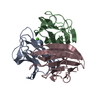


 PDBj
PDBj


 Assembly

 Type: D-saccharide, beta linking / Mass: 221.208 Da / Num. of mol.: 1

 Mass: 40.078 Da / Num. of mol.: 2 / Source method: obtained synthetically / Formula: Ca
 Type: L-peptide linking / Mass: 185.072 Da / Num. of mol.: 1 / Source method: obtained synthetically / Formula: C3H8NO6P
 Sample preparation
 / Beamline: ID14-2 / Wavelength: 0.933
  Processing