 Movie
Movie Controller


+ Open data

- Basic information
| Entry | Database: PDB / ID: 1pk6 | ||||||
|---|---|---|---|---|---|---|---|
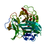
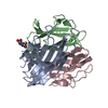
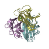
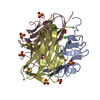
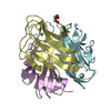
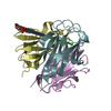
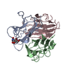
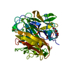
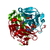
| Title | Globular Head of the Complement System Protein C1q | ||||||
 Components Components |
| ||||||
 Keywords Keywords |  IMMUNE SYSTEM / Complement system / C1q / immunology / jellyroll / IgG IMMUNE SYSTEM / Complement system / C1q / immunology / jellyroll / IgG | ||||||
| Function / homology |  Function and homology information Function and homology informationcomplement component C1 complex / complement component C1q complex / negative regulation of macrophage differentiation / synapse pruning / negative regulation of granulocyte differentiation / vertebrate eye-specific patterning / complement-mediated synapse pruning / collagen trimer / complement activation / Classical antibody-mediated complement activation ...complement component C1 complex / complement component C1q complex / negative regulation of macrophage differentiation / synapse pruning / negative regulation of granulocyte differentiation / vertebrate eye-specific patterning / complement-mediated synapse pruning / collagen trimer / complement activation / Classical antibody-mediated complement activation / neuron remodeling / Initial triggering of complement / complement activation, classical pathway / Regulation of Complement cascade / astrocyte activation / synapse organization / microglial cell activation / cell-cell signaling / amyloid-beta binding / postsynapse / collagen-containing extracellular matrix / blood microparticle / immune response / innate immune response / synapse / extracellular space / extracellular regionSimilarity search - Function | ||||||
| Biological species |  Homo sapiens (human) Homo sapiens (human) | ||||||
| Method | X-RAY DIFFRACTION / SYNCHROTRON / MOLECULAR REPLACEMENT / Resolution: 1.85 Å | ||||||
 Authors Authors | Gaboriaud, C. / Juanhuix, J. / Gruez, A. / Lacroix, M. / Darnault, C. / Pignol, D. / Verger, D. / Fontecilla-Camps, J.C. / Arlaud, G.J. | ||||||
 Citation Citation | Journal: J.Biol.Chem. / Year: 2003 Title: The crystal structure of the globular head of complement protein C1q provides a basis for its versatile recognition properties. Authors: Gaboriaud, C. / Juanhuix, J. / Gruez, A. / Lacroix, M. / Darnault, C. / Pignol, D. / Verger, D. / Fontecilla-Camps, J.C. / Arlaud, G.J. | ||||||
| History |
|
- Structure visualization
| Structure viewer | Molecule: MolmilJmol/JSmol |
|---|
- Downloads & links
-Download
| PDBx/mmCIF format | 1pk6.cif.gz | 94.4 KB | Display | PDBx/mmCIF format |
|---|---|---|---|---|
| PDB format | pdb1pk6.ent.gz | 71.1 KB | Display | PDB format |
| PDBx/mmJSON format | 1pk6.json.gz | Tree view | PDBx/mmJSON format | |
| Others |  Other downloads Other downloads |
-Validation report
| Arichive directory | https://data.pdbj.org/pub/pdb/validation_reports/pk/1pk6ftp://data.pdbj.org/pub/pdb/validation_reports/pk/1pk6 | HTTPS FTP |
|---|
-Related structure data
| Related structure data | 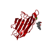 1gr3S S: Starting model for refinement |
|---|---|
| Similar structure data |


-Links
 PDBj
PDBj

- Assembly
| Deposited unit | 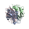
| ||||||||
|---|---|---|---|---|---|---|---|---|---|
| 1 |
| ||||||||
| Unit cell |
|
-Components
| #1: Protein | Mass: 14914.804 Da / Num. of mol.: 1 / Source method: isolated from a natural source / Details: serum / Source: (natural) Homo sapiens (human) / References: UniProt: P02745 |
|---|---|
| #2: Protein | Mass: 14996.058 Da / Num. of mol.: 1 / Source method: isolated from a natural source / Details: serum / Source: (natural) Homo sapiens (human) / References: UniProt: P02746 |
| #3: Protein | Mass: 14302.143 Da / Num. of mol.: 1 / Source method: isolated from a natural source / Details: serum / Source: (natural) Homo sapiens (human) / References: UniProt: P02747 |
| #4: Chemical | ChemComp-CA /   Mass: 40.078 Da / Num. of mol.: 1 / Source method: obtained synthetically / Formula: Ca Mass: 40.078 Da / Num. of mol.: 1 / Source method: obtained synthetically / Formula: Ca |
| #5: Water | ChemComp-HOH / Water Mass: 18.015 Da / Num. of mol.: 198 / Source method: isolated from a natural source / Formula: H2O Mass: 18.015 Da / Num. of mol.: 198 / Source method: isolated from a natural source / Formula: H2O |
-Experimental details
-Experiment
| Experiment | Method: X-RAY DIFFRACTION / Number of used crystals: 1 |
|---|
- Sample preparation
| Crystal | Density Matthews: 2.03 Å3/Da / Density % sol: 39.34 % | |||||||||||||||||||||||||||||||||||||||||||||||||||||||||||||||||||||||||||||
|---|---|---|---|---|---|---|---|---|---|---|---|---|---|---|---|---|---|---|---|---|---|---|---|---|---|---|---|---|---|---|---|---|---|---|---|---|---|---|---|---|---|---|---|---|---|---|---|---|---|---|---|---|---|---|---|---|---|---|---|---|---|---|---|---|---|---|---|---|---|---|---|---|---|---|---|---|---|---|
| Crystal grow | Temperature: 293 K / Method: vapor diffusion, hanging drop / pH: 7 Details: PEG 4000, CaCl2, B-mercaptoethanol, agarose, pH 7.0, VAPOR DIFFUSION, HANGING DROP, temperature 20K | |||||||||||||||||||||||||||||||||||||||||||||||||||||||||||||||||||||||||||||
| Crystal grow | *PLUS Temperature: 20 ℃ / pH: 7.6 / Method: vapor diffusion, hanging drop | |||||||||||||||||||||||||||||||||||||||||||||||||||||||||||||||||||||||||||||
| Components of the solutions | *PLUS
|
-Data collection
| Diffraction | Mean temperature: 100 K |
|---|---|
| Diffraction source | Source: SYNCHROTRON / Site: ESRF  / Beamline: ID29 / Wavelength: 1 Å / Beamline: ID29 / Wavelength: 1 Å |
| Detector | Type: ADSC / Detector: CCD / Date: Jan 28, 1998 |
| Radiation | Monochromator: SI(111) / Protocol: SINGLE WAVELENGTH / Monochromatic (M) / Laue (L): M / Scattering type: x-ray |
| Radiation wavelength | Wavelength: 1 Å / Relative weight: 1 |
| Reflection | Resolution: 1.85→25.4 Å / Num. all: 26237 / Num. obs: 26090 / % possible obs: 90 % / Redundancy: 4.73 % / Rmerge(I) obs: 0.073 / Net I/σ(I): 7.1 |
| Reflection shell | Resolution: 1.86→1.98 Å / Redundancy: 1.1 % / Rmerge(I) obs: 0.23 / Mean I/σ(I) obs: 1.7 / % possible all: 67.2 |
| Reflection | *PLUS Lowest resolution: 26 Å / Num. measured all: 116591 |
| Reflection shell | *PLUS Highest resolution: 1.85 Å / % possible obs: 67.2 % / Num. unique obs: 3614 / Num. measured obs: 3946 |
- Processing
| Software |
| ||||||||||||||||||||||||||||||||||||||||||||||||||||||||||||||||||||||||||||||||||||||||||||||||||||||||||||||||||||||||||||||||||
|---|---|---|---|---|---|---|---|---|---|---|---|---|---|---|---|---|---|---|---|---|---|---|---|---|---|---|---|---|---|---|---|---|---|---|---|---|---|---|---|---|---|---|---|---|---|---|---|---|---|---|---|---|---|---|---|---|---|---|---|---|---|---|---|---|---|---|---|---|---|---|---|---|---|---|---|---|---|---|---|---|---|---|---|---|---|---|---|---|---|---|---|---|---|---|---|---|---|---|---|---|---|---|---|---|---|---|---|---|---|---|---|---|---|---|---|---|---|---|---|---|---|---|---|---|---|---|---|---|---|---|---|
| Refinement | Method to determine structure: MOLECULAR REPLACEMENT Starting model: PDB: 1GR3 Resolution: 1.85→25.4 Å / Cor.coef. Fo:Fc: 0.95 / Cor.coef. Fo:Fc free: 0.932 / SU B: 5.396 / SU ML: 0.141 / Cross valid method: THROUGHOUT / ESU R: 0.25 / ESU R Free: 0.186 / Stereochemistry target values: MAXIMUM LIKELIHOOD Details: HYDROGENS HAVE BEEN ADDED IN THE RIDING POSITIONS. RESIDUES A90, A92, A160, B92, B93, B108, B109, B150, B163, B165, C89 HAVE DIFFUSE LATERAL CHAINS
| ||||||||||||||||||||||||||||||||||||||||||||||||||||||||||||||||||||||||||||||||||||||||||||||||||||||||||||||||||||||||||||||||||
| Solvent computation | Ion probe radii: 0.8 Å / Shrinkage radii: 0.8 Å / VDW probe radii: 1.4 Å / Solvent model: BABINET MODEL WITH MASK | ||||||||||||||||||||||||||||||||||||||||||||||||||||||||||||||||||||||||||||||||||||||||||||||||||||||||||||||||||||||||||||||||||
| Displacement parameters | Biso mean: 24.877 Å2
| ||||||||||||||||||||||||||||||||||||||||||||||||||||||||||||||||||||||||||||||||||||||||||||||||||||||||||||||||||||||||||||||||||
| Refinement step | Cycle: LAST / Resolution: 1.85→25.4 Å
| ||||||||||||||||||||||||||||||||||||||||||||||||||||||||||||||||||||||||||||||||||||||||||||||||||||||||||||||||||||||||||||||||||
| Refine LS restraints |
| ||||||||||||||||||||||||||||||||||||||||||||||||||||||||||||||||||||||||||||||||||||||||||||||||||||||||||||||||||||||||||||||||||
| LS refinement shell | Resolution: 1.851→1.898 Å / Total num. of bins used: 20 /
| ||||||||||||||||||||||||||||||||||||||||||||||||||||||||||||||||||||||||||||||||||||||||||||||||||||||||||||||||||||||||||||||||||
| Refinement | *PLUS Lowest resolution: 26 Å / Rfactor Rfree: 0.239 / Rfactor Rwork: 0.198 | ||||||||||||||||||||||||||||||||||||||||||||||||||||||||||||||||||||||||||||||||||||||||||||||||||||||||||||||||||||||||||||||||||
| Solvent computation | *PLUS | ||||||||||||||||||||||||||||||||||||||||||||||||||||||||||||||||||||||||||||||||||||||||||||||||||||||||||||||||||||||||||||||||||
| Displacement parameters | *PLUS | ||||||||||||||||||||||||||||||||||||||||||||||||||||||||||||||||||||||||||||||||||||||||||||||||||||||||||||||||||||||||||||||||||
| LS refinement shell | *PLUS Highest resolution: 1.85 Å / Lowest resolution: 1.98 Å / Rfactor Rfree: 0.27 / Rfactor Rwork: 0.28 |
