 Movie
Movie Controller


+ Open data

- Basic information
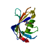
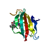
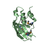
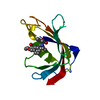
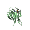
| Entry | Database: PDB / ID: 5ht1 | ||||||
|---|---|---|---|---|---|---|---|
| Title | Structure of apo C. glabrata FKBP12 | ||||||
 Components Components | FK506-binding protein 1 | ||||||
 Keywords Keywords |  ISOMERASE / FKBP12 / C. glabrata / pahtogenesis ISOMERASE / FKBP12 / C. glabrata / pahtogenesis | ||||||
| Function / homology |  Function and homology information Function and homology informationregulation of homoserine biosynthetic process / macrolide binding / peptidylprolyl isomerase / peptidyl-prolyl cis-trans isomerase activity / protein folding / chromatin organization / cytoplasmSimilarity search - Function | ||||||
| Biological species |  Candida glabrata (fungus) Candida glabrata (fungus) | ||||||
| Method | X-RAY DIFFRACTION / MOLECULAR REPLACEMENT / molecular replacement / Resolution: 2.651 Å | ||||||
 Authors Authors | Schumacher, M.A. | ||||||
 Citation Citation | Journal: Mbio / Year: 2016 Title: Structures of Pathogenic Fungal FKBP12s Reveal Possible Self-Catalysis Function. Authors: Tonthat, N.K. / Juvvadi, P.R. / Zhang, H. / Lee, S.C. / Venters, R. / Spicer, L. / Steinbach, W.J. / Heitman, J. / Schumacher, M.A. | ||||||
| History |
|
- Structure visualization
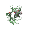
| Structure viewer | Molecule: MolmilJmol/JSmol |
|---|
- Downloads & links
-Download
| PDBx/mmCIF format | 5ht1.cif.gz | 33.9 KB | Display | PDBx/mmCIF format |
|---|---|---|---|---|
| PDB format | pdb5ht1.ent.gz | 22.4 KB | Display | PDB format |
| PDBx/mmJSON format | 5ht1.json.gz | Tree view | PDBx/mmJSON format | |
| Others |  Other downloads Other downloads |
-Validation report
| Arichive directory | https://data.pdbj.org/pub/pdb/validation_reports/ht/5ht1ftp://data.pdbj.org/pub/pdb/validation_reports/ht/5ht1 | HTTPS FTP |
|---|
-Related structure data
| Related structure data |  5htgC  5huaC  5hw6C 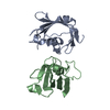 5hw7C 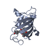 5hw8C  5hwbC  5hwcC  5i98C  5j6eC C: citing same article ( |
|---|---|
| Similar structure data |


-Links
 PDBj
PDBj

- Assembly
| Deposited unit | 
| ||||||||
|---|---|---|---|---|---|---|---|---|---|
| 1 |
| ||||||||
| Unit cell |
|
-Components
| #1: Protein | Mass: 12240.924 Da / Num. of mol.: 1 Source method: isolated from a genetically manipulated source Source: (gene. exp.) Candida glabrata (strain ATCC 2001 / CBS 138 / JCM 3761 / NBRC 0622 / NRRL Y-65) (fungus)Strain: ATCC 2001 / CBS 138 / JCM 3761 / NBRC 0622 / NRRL Y-65 Gene: FPR1, CAGL0K09724g / Production host:  Escherichia coli (E. coli) / References: UniProt: Q6FMA3, peptidylprolyl isomerase Escherichia coli (E. coli) / References: UniProt: Q6FMA3, peptidylprolyl isomerase |
|---|---|
| #2: Water | ChemComp-HOH / Water Mass: 18.015 Da / Num. of mol.: 20 / Source method: isolated from a natural source / Formula: H2O Mass: 18.015 Da / Num. of mol.: 20 / Source method: isolated from a natural source / Formula: H2O |
-Experimental details
-Experiment
| Experiment | Method: X-RAY DIFFRACTION / Number of used crystals: 1 |
|---|
- Sample preparation
| Crystal | Density Matthews: 2.59 Å3/Da / Density % sol: 52.56 % |
|---|---|
| Crystal grow | Temperature: 298 K / Method: vapor diffusion, hanging drop / Details: PEG 4000, 0.1 M Tris 7.5 |
-Data collection
| Diffraction | Mean temperature: 100 K |
|---|---|
| Diffraction source | Source: ROTATING ANODE / Type: OTHER / Wavelength: 1.5418 Å |
| Detector | Type: RIGAKU RAXIS HTC / Detector: IMAGE PLATE / Date: Mar 12, 2014 |
| Radiation | Protocol: SINGLE WAVELENGTH / Monochromatic (M) / Laue (L): M / Scattering type: x-ray |
| Radiation wavelength | Wavelength: 1.5418 Å / Relative weight: 1 |
| Reflection | Resolution: 2.65→43.508 Å / Num. obs: 3991 / % possible obs: 99.99 % / Redundancy: 3.3 % / Net I/σ(I): 11.8 |
-Phasing
| Phasing | Method: molecular replacement |
|---|
- Processing
| Software |
| ||||||||||||||||||||||||||||
|---|---|---|---|---|---|---|---|---|---|---|---|---|---|---|---|---|---|---|---|---|---|---|---|---|---|---|---|---|---|
| Refinement | Method to determine structure: MOLECULAR REPLACEMENT / Resolution: 2.651→43.508 Å / SU ML: 0.43 / Cross valid method: FREE R-VALUE / σ(F): 1.39 / Phase error: 23.58 / Stereochemistry target values: ML
| ||||||||||||||||||||||||||||
| Solvent computation | Shrinkage radii: 0.83 Å / VDW probe radii: 1.1 Å / Solvent model: FLAT BULK SOLVENT MODEL / Bsol: 21.584 Å2 / ksol: 0.378 e/Å3 | ||||||||||||||||||||||||||||
| Displacement parameters |
| ||||||||||||||||||||||||||||
| Refinement step | Cycle: LAST / Resolution: 2.651→43.508 Å
| ||||||||||||||||||||||||||||
| Refine LS restraints |
| ||||||||||||||||||||||||||||
| LS refinement shell |
|
