 Movie
Movie Controller

[English] 日本語
 Yorodumi
- PDB-3hx1: Crystal structure of the Slr1951 protein from Synechocystis sp. N... -
+ Open data

- Basic information
| Entry | Database: PDB / ID: 3hx1 | ||||||
|---|---|---|---|---|---|---|---|
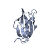
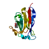
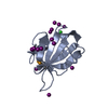
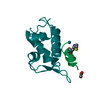
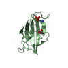
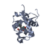
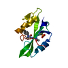
| Title | Crystal structure of the Slr1951 protein from Synechocystis sp. Northeast Structural Genomics Consortium Target SgR167A | ||||||
 Components Components | Slr1951 protein | ||||||
 Keywords Keywords |  Structural Genomics / Unknown function / P74513_SYNY3 / Slr1951 / Adenylate cyclase-like protein / NESG / SgR167A / PSI-2 / Protein Structure Initiative / Northeast Structural Genomics Consortium Structural Genomics / Unknown function / P74513_SYNY3 / Slr1951 / Adenylate cyclase-like protein / NESG / SgR167A / PSI-2 / Protein Structure Initiative / Northeast Structural Genomics Consortium | ||||||
| Function / homology |  Function and homology information Function and homology informationTumour Suppressor Smad4 - #20 / Tumour Suppressor Smad4 / Forkhead associated domain / Forkhead-associated (FHA) domain profile. / FHA domain / Forkhead-associated (FHA) domain / SMAD/FHA domain superfamily / Sandwich / Mainly BetaSimilarity search - Domain/homology | ||||||
| Biological species |  | ||||||
| Method | X-RAY DIFFRACTION / SYNCHROTRON / MAD / Resolution: 2.5 Å | ||||||
 Authors Authors | Vorobiev, S. / Chen, Y. / Seetharaman, J. / Janjua, J. / Xiao, R. / Ciccosanti, C. / Belote, R.L. / Everett, J.K. / Nair, R. / Acton, T.B. ...Vorobiev, S. / Chen, Y. / Seetharaman, J. / Janjua, J. / Xiao, R. / Ciccosanti, C. / Belote, R.L. / Everett, J.K. / Nair, R. / Acton, T.B. / Rost, B. / Montelione, G.T. / Hunt, J.F. / Tong, L. / Northeast Structural Genomics Consortium (NESG) | ||||||
 Citation Citation | Journal: To be Published Title: Crystal structure of the Slr1951 protein from Synechocystis sp. Authors: Vorobiev, S. / Chen, Y. / Seetharaman, J. / Janjua, J. / Xiao, R. / Ciccosanti, C. / Belote, R.L. / Everett, J.K. / Nair, R. / Acton, T.B. / Rost, B. / Montelione, G.T. / Hunt, J.F. / Tong, L. | ||||||
| History |
|
- Structure visualization
| Structure viewer | Molecule: MolmilJmol/JSmol |
|---|
- Downloads & links
-Download
| PDBx/mmCIF format | 3hx1.cif.gz | 52 KB | Display | PDBx/mmCIF format |
|---|---|---|---|---|
| PDB format | pdb3hx1.ent.gz | 40.4 KB | Display | PDB format |
| PDBx/mmJSON format | 3hx1.json.gz | Tree view | PDBx/mmJSON format | |
| Others |  Other downloads Other downloads |
-Validation report
| Arichive directory | https://data.pdbj.org/pub/pdb/validation_reports/hx/3hx1ftp://data.pdbj.org/pub/pdb/validation_reports/hx/3hx1 | HTTPS FTP |
|---|
-Related structure data
| Similar structure data | |
|---|---|
| Other databases |


-Links
 PDBj
PDBj

- Assembly
| Deposited unit | 
| ||||||||
|---|---|---|---|---|---|---|---|---|---|
| 1 | 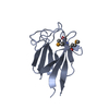
| ||||||||
| 2 | 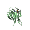
| ||||||||
| Unit cell |
| ||||||||
| Details | Monomer according to aggregation screening. |
-Components
| #1: Protein | Mass: 15224.563 Da / Num. of mol.: 2 / Fragment: residues 1-123 Source method: isolated from a genetically manipulated source Source: (gene. exp.) Escherichia coli (E. coli) / Strain (production host): BL21(DE3)+ Magic / References: UniProt: P74513#2: Water | ChemComp-HOH / | Water Mass: 18.015 Da / Num. of mol.: 46 / Source method: isolated from a natural source / Formula: H2O Mass: 18.015 Da / Num. of mol.: 46 / Source method: isolated from a natural source / Formula: H2O |
|---|
-Experimental details
-Experiment
| Experiment | Method: X-RAY DIFFRACTION / Number of used crystals: 1 |
|---|
- Sample preparation
| Crystal | Density Matthews: 2.17 Å3/Da / Density % sol: 43.37 % |
|---|---|
| Crystal grow | Temperature: 291 K / Method: microbatch under oil / pH: 8 Details: 40 % PEG 1000, 0.1M potassium phosphate monobasic, 0.1M TrisHCl, pH 8.0, Microbatch under oil, temperature 291K |
-Data collection
| Diffraction | Mean temperature: 100 K | ||||||||||||
|---|---|---|---|---|---|---|---|---|---|---|---|---|---|
| Diffraction source | Source: SYNCHROTRON / Site: SSRL  / Beamline: BL9-2 / Wavelength: 0.97931, 0.97907, 0.91162 / Beamline: BL9-2 / Wavelength: 0.97931, 0.97907, 0.91162 | ||||||||||||
| Detector | Type: MARMOSAIC 325 mm CCD / Detector: CCD / Date: Jun 14, 2009 | ||||||||||||
| Radiation | Protocol: MAD / Monochromatic (M) / Laue (L): M / Scattering type: x-ray | ||||||||||||
| Radiation wavelength |
| ||||||||||||
| Reflection | Resolution: 2.5→50 Å / Num. all: 17900 / Num. obs: 17793 / % possible obs: 99.4 % / Observed criterion σ(F): 0 / Observed criterion σ(I): 0 / Redundancy: 9.4 % / Biso Wilson estimate: 32 Å2 / Rmerge(I) obs: 0.141 / Net I/σ(I): 21.8 | ||||||||||||
| Reflection shell | Resolution: 2.5→2.59 Å / Redundancy: 5.4 % / Rmerge(I) obs: 0.629 / Mean I/σ(I) obs: 2.3 / Num. unique all: 1753 / % possible all: 99.6 |
- Processing
| Software |
| ||||||||||||||||||||||||||||||||
|---|---|---|---|---|---|---|---|---|---|---|---|---|---|---|---|---|---|---|---|---|---|---|---|---|---|---|---|---|---|---|---|---|---|
| Refinement | Method to determine structure: MAD / Resolution: 2.5→37.3 Å / Rfactor Rfree error: 0.009 / Data cutoff high absF: 234852.875 / Data cutoff low absF: 0 / Isotropic thermal model: RESTRAINED / Cross valid method: THROUGHOUT / σ(F): 1 / Stereochemistry target values: Engh & Huber
| ||||||||||||||||||||||||||||||||
| Solvent computation | Solvent model: FLAT MODEL / Bsol: 51.346 Å2 / ksol: 0.35 e/Å3 | ||||||||||||||||||||||||||||||||
| Displacement parameters | Biso mean: 54.5 Å2
| ||||||||||||||||||||||||||||||||
| Refine analyze |
| ||||||||||||||||||||||||||||||||
| Refinement step | Cycle: LAST / Resolution: 2.5→37.3 Å
| ||||||||||||||||||||||||||||||||
| Refine LS restraints |
| ||||||||||||||||||||||||||||||||
| LS refinement shell | Resolution: 2.5→2.66 Å / Rfactor Rfree error: 0.027 / Total num. of bins used: 6
| ||||||||||||||||||||||||||||||||
| Xplor file |
|
