 Movie
Movie Controller

[English] 日本語
 Yorodumi
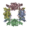
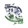
- PDB-5hoq: Apo structure of CalS11, TDP-rhamnose 3'-o-methyltransferase, an ... -
+ Open data

- Basic information
| Entry | Database: PDB / ID: 5hoq | ||||||||||||
|---|---|---|---|---|---|---|---|---|---|---|---|---|---|
| Title | Apo structure of CalS11, TDP-rhamnose 3'-o-methyltransferase, an enzyme in Calicheamicin biosynthesis | ||||||||||||
 Components Components | TDP-rhamnose 3'-O-methyltransferase (CalS11) | ||||||||||||
 Keywords Keywords |  TRANSFERASE / rhamnose methyltransferase Calicheamicin biosynthesis TRANSFERASE / rhamnose methyltransferase Calicheamicin biosynthesis | ||||||||||||
| Function / homology | Methyltransferase domain / Macrocin-O-methyltransferase / O-methyltransferase activity / Vaccinia Virus protein VP39 / S-adenosyl-L-methionine-dependent methyltransferase superfamily / Rossmann fold / 3-Layer(aba) Sandwich / Alpha Beta / CalS11 Function and homology information Function and homology information | ||||||||||||
| Biological species |  Micromonospora echinospora (bacteria) Micromonospora echinospora (bacteria) | ||||||||||||
| Method | X-RAY DIFFRACTION / SYNCHROTRON / MOLECULAR REPLACEMENT / Resolution: 1.793 Å | ||||||||||||
 Authors Authors | Han, L. / Helmich, K.E. / Singh, S. / Thorson, J.S. / Bingman, C.A. / Phillips Jr., G.N. / Enzyme Discovery for Natural Product Biosynthesis | ||||||||||||
 Citation Citation | Journal: Struct Dyn. / Year: 2016 Title: Loop dynamics of thymidine diphosphate-rhamnose 3'-O-methyltransferase (CalS11), an enzyme in calicheamicin biosynthesis. Authors: Han, L. / Singh, S. / Thorson, J.S. / Phillips, G.N. | ||||||||||||
| History |
|
- Structure visualization
| Structure viewer | Molecule: MolmilJmol/JSmol |
|---|
- Downloads & links
-Download
| PDBx/mmCIF format | 5hoq.cif.gz | 708.2 KB | Display | PDBx/mmCIF format |
|---|---|---|---|---|
| PDB format | pdb5hoq.ent.gz | 595.3 KB | Display | PDB format |
| PDBx/mmJSON format | 5hoq.json.gz | Tree view | PDBx/mmJSON format | |
| Others |  Other downloads Other downloads |
-Validation report
| Arichive directory | https://data.pdbj.org/pub/pdb/validation_reports/ho/5hoqftp://data.pdbj.org/pub/pdb/validation_reports/ho/5hoq | HTTPS FTP |
|---|
-Related structure data
| Related structure data |  4gf5S S: Starting model for refinement |
|---|---|
| Similar structure data |


-Links
 PDBj
PDBj- Assembly
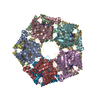
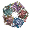
| Deposited unit | 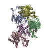
| |||||||||
|---|---|---|---|---|---|---|---|---|---|---|
| 1 | 
| |||||||||
| Unit cell |
| |||||||||
| Components on special symmetry positions |
|
-Components
| #1: Protein | Mass: 28950.844 Da / Num. of mol.: 5 Source method: isolated from a genetically manipulated source Source: (gene. exp.) Micromonospora echinospora (bacteria) / Production host: Escherichia coli (E. coli) / References: UniProt: Q8KNF1#2: Chemical | ChemComp-SO4 / Sulfate  Mass: 96.063 Da / Num. of mol.: 5 Mass: 96.063 Da / Num. of mol.: 5Source method: isolated from a genetically manipulated source Formula: SO4 #3: Water | ChemComp-HOH / | Water Mass: 18.015 Da / Num. of mol.: 1746 / Source method: isolated from a natural source / Formula: H2O Mass: 18.015 Da / Num. of mol.: 1746 / Source method: isolated from a natural source / Formula: H2O |
|---|
-Experimental details
-Experiment
| Experiment | Method: X-RAY DIFFRACTION / Number of used crystals: 1 |
|---|
- Sample preparation
| Crystal | Density Matthews: 2.81 Å3/Da / Density % sol: 56.2 % |
|---|---|
| Crystal grow | Temperature: 293 K / Method: vapor diffusion, hanging drop / pH: 7.5 Details: 1:1 ratio mixture of 16mg/ml protein and reservoir solution (21% PEG3350, 0.1M LiSO4, 0.1M HEPES pH 7.5), VAPOR DIFFUSION, HANGING DROP, temperature 293K |
-Data collection
| Diffraction | Mean temperature: 100 K |
|---|---|
| Diffraction source | Source: SYNCHROTRON / Site: APS  / Beamline: 21-ID-F / Wavelength: 0.97857 Å / Beamline: 21-ID-F / Wavelength: 0.97857 Å |
| Detector | Type: MARMOSAIC 225 mm CCD / Detector: CCD / Date: Feb 2, 2013 |
| Radiation | Protocol: SINGLE WAVELENGTH / Monochromatic (M) / Laue (L): M / Scattering type: x-ray |
| Radiation wavelength | Wavelength: 0.97857 Å / Relative weight: 1 |
| Reflection | Resolution: 1.793→47.888 Å / Num. obs: 147725 / % possible obs: 99 % / Redundancy: 7.4 % / CC1/2: 0.997 / Rmerge(I) obs: 0.1316 / Net I/σ(I): 10.25 |
| Reflection shell | Resolution: 1.793→1.86 Å / Redundancy: 6.8 % / Rmerge(I) obs: 1.234 / Mean I/σ(I) obs: 1.39 / % possible all: 90 |
- Processing
| Software |
| |||||||||||||||||||||||||||||||||||||||||||||||||||||||||||||||||||||||||||||||||||||||||||||||||||||||||||||||||||||||||||||||||||||||||||||||||||||||||||||||||||||||||||||||||||||||||||||||||||||||||||||||||||||||||
|---|---|---|---|---|---|---|---|---|---|---|---|---|---|---|---|---|---|---|---|---|---|---|---|---|---|---|---|---|---|---|---|---|---|---|---|---|---|---|---|---|---|---|---|---|---|---|---|---|---|---|---|---|---|---|---|---|---|---|---|---|---|---|---|---|---|---|---|---|---|---|---|---|---|---|---|---|---|---|---|---|---|---|---|---|---|---|---|---|---|---|---|---|---|---|---|---|---|---|---|---|---|---|---|---|---|---|---|---|---|---|---|---|---|---|---|---|---|---|---|---|---|---|---|---|---|---|---|---|---|---|---|---|---|---|---|---|---|---|---|---|---|---|---|---|---|---|---|---|---|---|---|---|---|---|---|---|---|---|---|---|---|---|---|---|---|---|---|---|---|---|---|---|---|---|---|---|---|---|---|---|---|---|---|---|---|---|---|---|---|---|---|---|---|---|---|---|---|---|---|---|---|---|---|---|---|---|---|---|---|---|---|---|---|---|---|---|---|---|
| Refinement | Method to determine structure: MOLECULAR REPLACEMENT Starting model: 4GF5 Resolution: 1.793→47.888 Å / SU ML: 0.21 / Cross valid method: FREE R-VALUE / σ(F): 1.34 / Phase error: 18.06 / Stereochemistry target values: ML
| |||||||||||||||||||||||||||||||||||||||||||||||||||||||||||||||||||||||||||||||||||||||||||||||||||||||||||||||||||||||||||||||||||||||||||||||||||||||||||||||||||||||||||||||||||||||||||||||||||||||||||||||||||||||||
| Solvent computation | Shrinkage radii: 0.9 Å / VDW probe radii: 1.11 Å / Solvent model: FLAT BULK SOLVENT MODEL | |||||||||||||||||||||||||||||||||||||||||||||||||||||||||||||||||||||||||||||||||||||||||||||||||||||||||||||||||||||||||||||||||||||||||||||||||||||||||||||||||||||||||||||||||||||||||||||||||||||||||||||||||||||||||
| Refinement step | Cycle: LAST / Resolution: 1.793→47.888 Å
| |||||||||||||||||||||||||||||||||||||||||||||||||||||||||||||||||||||||||||||||||||||||||||||||||||||||||||||||||||||||||||||||||||||||||||||||||||||||||||||||||||||||||||||||||||||||||||||||||||||||||||||||||||||||||
| Refine LS restraints |
| |||||||||||||||||||||||||||||||||||||||||||||||||||||||||||||||||||||||||||||||||||||||||||||||||||||||||||||||||||||||||||||||||||||||||||||||||||||||||||||||||||||||||||||||||||||||||||||||||||||||||||||||||||||||||
| LS refinement shell |
| |||||||||||||||||||||||||||||||||||||||||||||||||||||||||||||||||||||||||||||||||||||||||||||||||||||||||||||||||||||||||||||||||||||||||||||||||||||||||||||||||||||||||||||||||||||||||||||||||||||||||||||||||||||||||
| Refinement TLS params. | Method: refined / Refine-ID: X-RAY DIFFRACTION
| |||||||||||||||||||||||||||||||||||||||||||||||||||||||||||||||||||||||||||||||||||||||||||||||||||||||||||||||||||||||||||||||||||||||||||||||||||||||||||||||||||||||||||||||||||||||||||||||||||||||||||||||||||||||||
| Refinement TLS group |
|
