 Movie
Movie Controller


+ Open data

- Basic information
| Entry | Database: PDB / ID: 4gf5 | ||||||
|---|---|---|---|---|---|---|---|
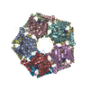
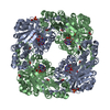
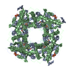
| Title | Crystal Structure of Calicheamicin Methyltransferase, CalS11 | ||||||
 Components Components | CalS11 | ||||||
 Keywords Keywords |  TRANSFERASE / CalS11 / SAH / Methyltransferase / Calicheamicin / Structural Genomics / Protein Structure Initiative / PSI / PSI-Biology / NATPRO / Enzyme Discovery for Natural Products Biosynthesis / Center for Eukaryotic Structural Genomics / CESG / Enzyme Discovery for Natural Product Biosynthesis TRANSFERASE / CalS11 / SAH / Methyltransferase / Calicheamicin / Structural Genomics / Protein Structure Initiative / PSI / PSI-Biology / NATPRO / Enzyme Discovery for Natural Products Biosynthesis / Center for Eukaryotic Structural Genomics / CESG / Enzyme Discovery for Natural Product Biosynthesis | ||||||
| Function / homology |  Function and homology information Function and homology information | ||||||
| Biological species |  Micromonospora echinospora (bacteria) Micromonospora echinospora (bacteria) | ||||||
| Method | X-RAY DIFFRACTION / SYNCHROTRON / MOLECULAR REPLACEMENT / molecular replacement / Resolution: 2.2 Å | ||||||
 Authors Authors | Helmich, K.E. / Singh, S. / Thorson, J.S. / Phillips Jr., G.N. / Enzyme Discovery for Natural Product Biosynthesis (NatPro) / Center for Eukaryotic Structural Genomics (CESG) | ||||||
 Citation Citation | Journal: to be published | ||||||
| History |
|
- Structure visualization
| Structure viewer | Molecule: MolmilJmol/JSmol |
|---|
- Downloads & links
-Download
| PDBx/mmCIF format | 4gf5.cif.gz | 1 MB | Display | PDBx/mmCIF format |
|---|---|---|---|---|
| PDB format | pdb4gf5.ent.gz | 860.1 KB | Display | PDB format |
| PDBx/mmJSON format | 4gf5.json.gz | Tree view | PDBx/mmJSON format | |
| Others |  Other downloads Other downloads |
-Validation report
| Arichive directory | https://data.pdbj.org/pub/pdb/validation_reports/gf/4gf5ftp://data.pdbj.org/pub/pdb/validation_reports/gf/4gf5 | HTTPS FTP |
|---|
-Related structure data
| Related structure data |  3tosS S: Starting model for refinement |
|---|---|
| Similar structure data | |
| Other databases |


-Links
 PDBj
PDBj- Assembly
| Deposited unit | 
| ||||||||
|---|---|---|---|---|---|---|---|---|---|
| 1 | 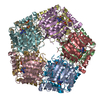
| ||||||||
| 2 | 
| ||||||||
| Unit cell |
| ||||||||
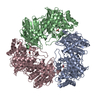
| Details | The biological unit is a decamer. There are 2 biological units in the asymmetric unit (chains A-J and chains K-T) |
-Components
| #1: Protein | Mass: 28950.844 Da / Num. of mol.: 20 Source method: isolated from a genetically manipulated source Source: (gene. exp.) Micromonospora echinospora (bacteria) / Strain: Micromonospora echinospora / Gene: AAM70337, calS11 / Plasmid: pET28a / Production host: Escherichia coli (E. coli) / Strain (production host): B834 / References: UniProt: Q8KNF1#2: Chemical | ChemComp-SAH / S-Adenosyl-L-homocysteine  Type: L-peptide linking / Mass: 384.411 Da / Num. of mol.: 20 / Source method: obtained synthetically / Formula: C14H20N6O5S Type: L-peptide linking / Mass: 384.411 Da / Num. of mol.: 20 / Source method: obtained synthetically / Formula: C14H20N6O5S#3: Chemical | ChemComp-SO4 / Sulfate  Mass: 96.063 Da / Num. of mol.: 28 / Source method: obtained synthetically / Formula: SO4 Mass: 96.063 Da / Num. of mol.: 28 / Source method: obtained synthetically / Formula: SO4#4: Water | ChemComp-HOH / | Water Mass: 18.015 Da / Num. of mol.: 2734 / Source method: isolated from a natural source / Formula: H2O Mass: 18.015 Da / Num. of mol.: 2734 / Source method: isolated from a natural source / Formula: H2O |
|---|
-Experimental details
-Experiment
| Experiment | Method: X-RAY DIFFRACTION / Number of used crystals: 1 |
|---|
- Sample preparation
| Crystal | Density Matthews: 2.35 Å3/Da / Density % sol: 47.65 % |
|---|---|
| Crystal grow | Temperature: 298 K / Method: vapor diffusion, hanging drop / pH: 8.5 Details: Protein solution (11 mg/ml CalS11, 25mM Tris pH 8.0, 5 mg/ml calicheamicin) mixed in a 1:1 ratio with the well solution (25% PEG 3350, .2M LiSO4, .1M Tris pH 8.5) Cryoprotected with 30% PEG ...Details: Protein solution (11 mg/ml CalS11, 25mM Tris pH 8.0, 5 mg/ml calicheamicin) mixed in a 1:1 ratio with the well solution (25% PEG 3350, .2M LiSO4, .1M Tris pH 8.5) Cryoprotected with 30% PEG 3350, .2M LiSO4, .1M Tris pH 8.5, Vapor diffusion, hanging drop, temperature 298K |
-Data collection
| Diffraction | Mean temperature: 100 K | |||||||||||||||||||||||||||||||||||||||||||||||||||||||||||||||||||||||||||||||||||||||||||||||||||||||||||||||||||||||||||||||||||||||||||||||||||
|---|---|---|---|---|---|---|---|---|---|---|---|---|---|---|---|---|---|---|---|---|---|---|---|---|---|---|---|---|---|---|---|---|---|---|---|---|---|---|---|---|---|---|---|---|---|---|---|---|---|---|---|---|---|---|---|---|---|---|---|---|---|---|---|---|---|---|---|---|---|---|---|---|---|---|---|---|---|---|---|---|---|---|---|---|---|---|---|---|---|---|---|---|---|---|---|---|---|---|---|---|---|---|---|---|---|---|---|---|---|---|---|---|---|---|---|---|---|---|---|---|---|---|---|---|---|---|---|---|---|---|---|---|---|---|---|---|---|---|---|---|---|---|---|---|---|---|---|---|
| Diffraction source | Source: SYNCHROTRON / Site: APS  / Beamline: 21-ID-F / Wavelength: 0.97872 Å / Beamline: 21-ID-F / Wavelength: 0.97872 Å | |||||||||||||||||||||||||||||||||||||||||||||||||||||||||||||||||||||||||||||||||||||||||||||||||||||||||||||||||||||||||||||||||||||||||||||||||||
| Detector | Type: MARMOSAIC 225 mm CCD / Detector: CCD / Date: Nov 5, 2011 | |||||||||||||||||||||||||||||||||||||||||||||||||||||||||||||||||||||||||||||||||||||||||||||||||||||||||||||||||||||||||||||||||||||||||||||||||||
| Radiation | Monochromator: C(111) / Protocol: SINGLE WAVELENGTH / Scattering type: x-ray | |||||||||||||||||||||||||||||||||||||||||||||||||||||||||||||||||||||||||||||||||||||||||||||||||||||||||||||||||||||||||||||||||||||||||||||||||||
| Radiation wavelength | Wavelength: 0.97872 Å / Relative weight: 1 | |||||||||||||||||||||||||||||||||||||||||||||||||||||||||||||||||||||||||||||||||||||||||||||||||||||||||||||||||||||||||||||||||||||||||||||||||||
| Reflection | Resolution: 2.2→50 Å / Num. all: 254196 / Num. obs: 254196 / % possible obs: 94.8 % / Observed criterion σ(F): 0 / Observed criterion σ(I): 0 / Redundancy: 3.9 % / Rmerge(I) obs: 0.11 / Χ2: 0.797 / Net I/σ(I): 6.3 | |||||||||||||||||||||||||||||||||||||||||||||||||||||||||||||||||||||||||||||||||||||||||||||||||||||||||||||||||||||||||||||||||||||||||||||||||||
| Reflection shell |
|
-Phasing
| Phasing | Method: molecular replacement | |||||||||
|---|---|---|---|---|---|---|---|---|---|---|
| Phasing MR |
|
- Processing
| Software |
| |||||||||||||||||||||||||||||||||||||||||||||||||||||||||||||||||||||||||||||||||||||||||||||||||||||||||
|---|---|---|---|---|---|---|---|---|---|---|---|---|---|---|---|---|---|---|---|---|---|---|---|---|---|---|---|---|---|---|---|---|---|---|---|---|---|---|---|---|---|---|---|---|---|---|---|---|---|---|---|---|---|---|---|---|---|---|---|---|---|---|---|---|---|---|---|---|---|---|---|---|---|---|---|---|---|---|---|---|---|---|---|---|---|---|---|---|---|---|---|---|---|---|---|---|---|---|---|---|---|---|---|---|---|---|
| Refinement | Method to determine structure: MOLECULAR REPLACEMENT Starting model: pdb entry 3TOS Resolution: 2.2→49.322 Å / Occupancy max: 1 / Occupancy min: 0 / FOM work R set: 0.8689 / SU ML: 0.25 / σ(F): 0 / Phase error: 20.93 / Stereochemistry target values: ML
| |||||||||||||||||||||||||||||||||||||||||||||||||||||||||||||||||||||||||||||||||||||||||||||||||||||||||
| Solvent computation | Shrinkage radii: 0.9 Å / VDW probe radii: 1.11 Å / Solvent model: FLAT BULK SOLVENT MODEL | |||||||||||||||||||||||||||||||||||||||||||||||||||||||||||||||||||||||||||||||||||||||||||||||||||||||||
| Displacement parameters | Biso max: 85.75 Å2 / Biso mean: 15.2599 Å2 / Biso min: 1.83 Å2 | |||||||||||||||||||||||||||||||||||||||||||||||||||||||||||||||||||||||||||||||||||||||||||||||||||||||||
| Refinement step | Cycle: LAST / Resolution: 2.2→49.322 Å
| |||||||||||||||||||||||||||||||||||||||||||||||||||||||||||||||||||||||||||||||||||||||||||||||||||||||||
| Refine LS restraints |
| |||||||||||||||||||||||||||||||||||||||||||||||||||||||||||||||||||||||||||||||||||||||||||||||||||||||||
| LS refinement shell | Refine-ID: X-RAY DIFFRACTION / Total num. of bins used: 14
|
