 Movie
Movie Controller


+ Open data

- Basic information
| Entry | Database: PDB / ID: 4y4l | ||||||
|---|---|---|---|---|---|---|---|
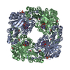
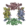
| Title | Crystal structure of yeast Thi4-C205S | ||||||
 Components Components | Thiamine thiazole synthase | ||||||
 Keywords Keywords | BIOSYNTHETIC PROTEIN | ||||||
| Function / homology |  Function and homology information Function and homology informationcysteine-dependent adenosine diphosphate thiazole synthase / cysteine-dependent adenosine diphosphate thiazole synthase activity / thiazole biosynthetic process / thiamine biosynthetic process / : / ferrous iron binding / iron ion binding / nucleus / cytosol Similarity search - Function | ||||||
| Biological species |  | ||||||
| Method |  X-RAY DIFFRACTION / SYNCHROTRON / MOLECULAR REPLACEMENT / Resolution: 2 Å X-RAY DIFFRACTION / SYNCHROTRON / MOLECULAR REPLACEMENT / Resolution: 2 Å | ||||||
 Authors Authors | Zhang, X. / Ealick, S.E. | ||||||
 Citation Citation | Journal: Biochemistry / Year: 2016 Title: Structural Basis for Iron-Mediated Sulfur Transfer in Archael and Yeast Thiazole Synthases. Authors: Zhang, X. / Eser, B.E. / Chanani, P.K. / Begley, T.P. / Ealick, S.E. | ||||||
| History |
|
- Structure visualization
| Structure viewer | Molecule: MolmilJmol/JSmol |
|---|
- Downloads & links
-Download
| PDBx/mmCIF format | 4y4l.cif.gz | 243.9 KB | Display | PDBx/mmCIF format |
|---|---|---|---|---|
| PDB format | pdb4y4l.ent.gz | 196.5 KB | Display | PDB format |
| PDBx/mmJSON format | 4y4l.json.gz | Tree view | PDBx/mmJSON format | |
| Others |  Other downloads Other downloads |
-Validation report
| Summary document | 4y4l_validation.pdf.gz | 1.4 MB | Display | wwPDB validaton report |
|---|---|---|---|---|
| Full document | 4y4l_full_validation.pdf.gz | 1.4 MB | Display | |
| Data in XML | 4y4l_validation.xml.gz | 45.7 KB | Display | |
| Data in CIF | 4y4l_validation.cif.gz | 63.6 KB | Display | |
| Arichive directory | https://data.pdbj.org/pub/pdb/validation_reports/y4/4y4lftp://data.pdbj.org/pub/pdb/validation_reports/y4/4y4l | HTTPS FTP |
-Related structure data
| Related structure data |  4y4mC  4y4nC  3fpzS S: Starting model for refinement C: citing same article ( |
|---|---|
| Similar structure data |


-Links
 PDBj
PDBj- Assembly
| Deposited unit | 
| ||||||||
|---|---|---|---|---|---|---|---|---|---|
| 1 | 
| ||||||||
| 2 | 
| ||||||||
| Unit cell |
|
-Components
| #1: Protein | Mass: 37185.180 Da / Num. of mol.: 4 / Mutation: C205S Source method: isolated from a genetically manipulated source Source: (gene. exp.) Strain: ATCC 204508 / S288c / Gene: THI4, ESP35, MOL1, YGR144W, G6620 / Production host:  #2: Chemical | ChemComp-48N / (   Mass: 598.352 Da / Num. of mol.: 4 / Source method: obtained synthetically / Formula: C17H24N6O14P2 Mass: 598.352 Da / Num. of mol.: 4 / Source method: obtained synthetically / Formula: C17H24N6O14P2#3: Water | ChemComp-HOH / |  Mass: 18.015 Da / Num. of mol.: 333 / Source method: isolated from a natural source / Formula: H2O Mass: 18.015 Da / Num. of mol.: 333 / Source method: isolated from a natural source / Formula: H2O |
|---|
-Experimental details
-Experiment
| Experiment | Method: X-RAY DIFFRACTION |
|---|
- Sample preparation
| Crystal | Density Matthews: 2.1 Å3/Da / Density % sol: 41.5 % |
|---|---|
| Crystal grow | Temperature: 295 K / Method: evaporation / pH: 8.5 Details: 25% (w/v) PEG1500, 0.0125 M succinic acid, 0.04375M sodium dihydrogen phosphate, and 0.04375M glycine, pH 8.5 |
-Data collection
| Diffraction | Mean temperature: 100 K |
|---|---|
| Diffraction source | Source: SYNCHROTRON / Site: APS  / Beamline: 24-ID-C / Wavelength: 0.9791 Å / Beamline: 24-ID-C / Wavelength: 0.9791 Å |
| Detector | Type: ADSC QUANTUM 315 / Detector: CCD / Date: Aug 19, 2011 |
| Radiation | Protocol: SINGLE WAVELENGTH / Monochromatic (M) / Laue (L): M / Scattering type: x-ray |
| Radiation wavelength | Wavelength: 0.9791 Å / Relative weight: 1 |
| Reflection | Resolution: 2→40 Å / Num. obs: 78765 / % possible obs: 100 % / Redundancy: 5 % / Rsym value: 0.07 / Net I/σ(I): 14.5 |
- Processing
| Software |
| ||||||||||||||||||||||||||||||||||||||||||||||||||||||||||||||||||||||||||||||||||||||||||||||||||||||||||||||||||||||||||||||||||||||||||||||||||||||||||||||||||||||||||||||||||||||
|---|---|---|---|---|---|---|---|---|---|---|---|---|---|---|---|---|---|---|---|---|---|---|---|---|---|---|---|---|---|---|---|---|---|---|---|---|---|---|---|---|---|---|---|---|---|---|---|---|---|---|---|---|---|---|---|---|---|---|---|---|---|---|---|---|---|---|---|---|---|---|---|---|---|---|---|---|---|---|---|---|---|---|---|---|---|---|---|---|---|---|---|---|---|---|---|---|---|---|---|---|---|---|---|---|---|---|---|---|---|---|---|---|---|---|---|---|---|---|---|---|---|---|---|---|---|---|---|---|---|---|---|---|---|---|---|---|---|---|---|---|---|---|---|---|---|---|---|---|---|---|---|---|---|---|---|---|---|---|---|---|---|---|---|---|---|---|---|---|---|---|---|---|---|---|---|---|---|---|---|---|---|---|---|
| Refinement | Method to determine structure: MOLECULAR REPLACEMENT Starting model: 3FPZ Resolution: 2→38.27 Å / Cor.coef. Fo:Fc: 0.957 / Cor.coef. Fo:Fc free: 0.947 / Cross valid method: THROUGHOUT / ESU R: 0.18 / ESU R Free: 0.141 / Stereochemistry target values: MAXIMUM LIKELIHOOD / Details: HYDROGENS HAVE BEEN ADDED IN THE RIDING POSITIONS
| ||||||||||||||||||||||||||||||||||||||||||||||||||||||||||||||||||||||||||||||||||||||||||||||||||||||||||||||||||||||||||||||||||||||||||||||||||||||||||||||||||||||||||||||||||||||
| Solvent computation | Ion probe radii: 0.8 Å / Shrinkage radii: 0.8 Å / VDW probe radii: 1.4 Å / Solvent model: MASK | ||||||||||||||||||||||||||||||||||||||||||||||||||||||||||||||||||||||||||||||||||||||||||||||||||||||||||||||||||||||||||||||||||||||||||||||||||||||||||||||||||||||||||||||||||||||
| Displacement parameters | Biso mean: 23.019 Å2
| ||||||||||||||||||||||||||||||||||||||||||||||||||||||||||||||||||||||||||||||||||||||||||||||||||||||||||||||||||||||||||||||||||||||||||||||||||||||||||||||||||||||||||||||||||||||
| Refinement step | Cycle: LAST / Resolution: 2→38.27 Å
| ||||||||||||||||||||||||||||||||||||||||||||||||||||||||||||||||||||||||||||||||||||||||||||||||||||||||||||||||||||||||||||||||||||||||||||||||||||||||||||||||||||||||||||||||||||||
| Refine LS restraints |
|