 Movie
Movie Controller

[English] 日本語
 Yorodumi
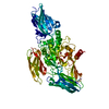
- PDB-5h67: Crystal structure of the Bacillus subtilis SMC head domain comple... -
+ Open data

- Basic information
| Entry | Database: PDB / ID: 5h67 | ||||||
|---|---|---|---|---|---|---|---|
| Title | Crystal structure of the Bacillus subtilis SMC head domain complexed with the cognate ScpA C-terminal domain and soaked ATP | ||||||
 Components Components |
| ||||||
 Keywords Keywords | DNA BINDING PROTEIN/CELL CYCLE /  SMC protein / DNA BINDING PROTEIN-CELL CYCLE complex SMC protein / DNA BINDING PROTEIN-CELL CYCLE complex | ||||||
| Function / homology |  Function and homology information Function and homology informationchromosome condensation / sister chromatid cohesion / chromosome segregation / chromosome / DNA replication / cell division / ATP hydrolysis activity / DNA binding / ATP binding / identical protein binding / cytoplasmSimilarity search - Function | ||||||
| Biological species |  Bacillus subtilis (bacteria) Bacillus subtilis (bacteria) | ||||||
| Method | X-RAY DIFFRACTION / SYNCHROTRON / MOLECULAR REPLACEMENT / Resolution: 2.07203506543 Å | ||||||
 Authors Authors | Kamada, K. / Hirano, T. | ||||||
| Funding support |  Japan, 1items Japan, 1items
| ||||||
 Citation Citation | Journal: Structure / Year: 2017 Title: Overall Shapes of the SMC-ScpAB Complex Are Determined by Balance between Constraint and Relaxation of Its Structural Parts Authors: Kamada, K. / Su'etsugu, M. / Takada, H. / Miyata, M. / Hirano, T. | ||||||
| History |
|
- Structure visualization
| Structure viewer | Molecule: MolmilJmol/JSmol |
|---|
- Downloads & links
-Download
| PDBx/mmCIF format | 5h67.cif.gz | 133.9 KB | Display | PDBx/mmCIF format |
|---|---|---|---|---|
| PDB format | pdb5h67.ent.gz | 82.7 KB | Display | PDB format |
| PDBx/mmJSON format | 5h67.json.gz | Tree view | PDBx/mmJSON format | |
| Others |  Other downloads Other downloads |
-Validation report
| Arichive directory | https://data.pdbj.org/pub/pdb/validation_reports/h6/5h67ftp://data.pdbj.org/pub/pdb/validation_reports/h6/5h67 | HTTPS FTP |
|---|
-Related structure data
| Related structure data | 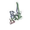 5h66SC 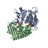 5h68C  5h69C S: Starting model for refinement C: citing same article ( |
|---|---|
| Similar structure data |


-Links
 PDBj
PDBj- Assembly
| Deposited unit | 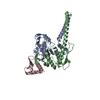
| ||||||||||||
|---|---|---|---|---|---|---|---|---|---|---|---|---|---|
| 1 |
| ||||||||||||
| Unit cell |
|
-Components
| #1: Protein | Mass: 22517.609 Da / Num. of mol.: 1 / Fragment: N-terminal, UNP residues 1-199 Source method: isolated from a genetically manipulated source Source: (gene. exp.) Bacillus subtilis (strain 168) (bacteria)Strain: 168 / Gene: smc, ylqA, BSU15940 / Plasmid: pET-22b / Details (production host): coexpression / Production host: Escherichia coli BL21(DE3) (bacteria) / Strain (production host): BL21(DE3) / References: UniProt: P51834 |
|---|---|
| #2: Protein | Mass: 21466.559 Da / Num. of mol.: 1 / Fragment: C-terminal, UNP residues 1000-1186 / Mutation: E1118Q Source method: isolated from a genetically manipulated source Source: (gene. exp.) Bacillus subtilis (strain 168) (bacteria)Strain: 168 / Gene: smc, ylqA, BSU15940 / Plasmid: pET-22b / Details (production host): coexpression / Production host: Escherichia coli BL21(DE3) (bacteria) / Strain (production host): BL21(DE3) / References: UniProt: P51834 |
| #3: Protein | Mass: 9289.652 Da / Num. of mol.: 1 / Fragment: UNP residues 176-251 Source method: isolated from a genetically manipulated source Source: (gene. exp.) Bacillus subtilis (strain 168) (bacteria)Strain: 168 / Gene: scpA, ypuG, BSU23220 / Plasmid: pET-28m / Details (production host): modified / Production host: Escherichia coli BL21(DE3) (bacteria) / Strain (production host): BL21(DE3) / References: UniProt: P35154 |
| #4: Chemical | ChemComp-ATP / Adenosine triphosphate  Mass: 507.181 Da / Num. of mol.: 1 / Source method: obtained synthetically / Formula: C10H16N5O13P3 / Comment: ATP, energy-carrying molecule*YM Mass: 507.181 Da / Num. of mol.: 1 / Source method: obtained synthetically / Formula: C10H16N5O13P3 / Comment: ATP, energy-carrying molecule*YM |
| #5: Water | ChemComp-HOH / Water Mass: 18.015 Da / Num. of mol.: 183 / Source method: isolated from a natural source / Formula: H2O Mass: 18.015 Da / Num. of mol.: 183 / Source method: isolated from a natural source / Formula: H2O |
-Experimental details
-Experiment
| Experiment | Method: X-RAY DIFFRACTION / Number of used crystals: 1 |
|---|
- Sample preparation
| Crystal | Density Matthews: 3.67 Å3/Da / Density % sol: 66.46 % / Description: Orthogonal plate |
|---|---|
| Crystal grow | Temperature: 277 K / Method: vapor diffusion, hanging drop / pH: 7.4 Details: 3.5M sodium formate (pH 7.4), the crystals were soaked in the reservoir solution containing 10mM Mg-ATP |
-Data collection
| Diffraction | Mean temperature: 100 K |
|---|---|
| Diffraction source | Source: SYNCHROTRON / Site: SPring-8 / Beamline: BL26B1 / Wavelength: 1 Å |
| Detector | Type: MARMOSAIC 225 mm CCD / Detector: CCD / Date: Feb 1, 2012 |
| Radiation | Monochromator: SI 111 / Protocol: SINGLE WAVELENGTH / Monochromatic (M) / Laue (L): M / Scattering type: x-ray |
| Radiation wavelength | Wavelength: 1 Å / Relative weight: 1 |
| Reflection | Resolution: 2.07→50 Å / Num. obs: 44563 / % possible obs: 98.4 % / Observed criterion σ(F): 0 / Observed criterion σ(I): -3 / Redundancy: 3.5 % / Biso Wilson estimate: 32.0993476008 Å2 / Rmerge(I) obs: 0.039 / Rsym value: 0.039 / Net I/av σ(I): 27.5 / Net I/σ(I): 14.8 |
| Reflection shell | Resolution: 2.07→2.11 Å / Redundancy: 2.9 % / Rmerge(I) obs: 0.244 / Rsym value: 0.244 / % possible all: 86.7 |
- Processing
| Software |
| |||||||||||||||||||||||||||||||||||||||||||||||||||||||||||||||||||||||||||||||||||||||||||||||||||||||||||||||||||||||
|---|---|---|---|---|---|---|---|---|---|---|---|---|---|---|---|---|---|---|---|---|---|---|---|---|---|---|---|---|---|---|---|---|---|---|---|---|---|---|---|---|---|---|---|---|---|---|---|---|---|---|---|---|---|---|---|---|---|---|---|---|---|---|---|---|---|---|---|---|---|---|---|---|---|---|---|---|---|---|---|---|---|---|---|---|---|---|---|---|---|---|---|---|---|---|---|---|---|---|---|---|---|---|---|---|---|---|---|---|---|---|---|---|---|---|---|---|---|---|---|---|
| Refinement | Method to determine structure: MOLECULAR REPLACEMENT Starting model: 5H66 Resolution: 2.07203506543→40.627174663 Å / SU ML: 0.199736653153 / Cross valid method: FREE R-VALUE / σ(F): 1.36326163552 / Phase error: 22.5281067016
| |||||||||||||||||||||||||||||||||||||||||||||||||||||||||||||||||||||||||||||||||||||||||||||||||||||||||||||||||||||||
| Solvent computation | Shrinkage radii: 0.9 Å / VDW probe radii: 1.11 Å | |||||||||||||||||||||||||||||||||||||||||||||||||||||||||||||||||||||||||||||||||||||||||||||||||||||||||||||||||||||||
| Displacement parameters | Biso mean: 44.2498877626 Å2 | |||||||||||||||||||||||||||||||||||||||||||||||||||||||||||||||||||||||||||||||||||||||||||||||||||||||||||||||||||||||
| Refinement step | Cycle: LAST / Resolution: 2.07203506543→40.627174663 Å
| |||||||||||||||||||||||||||||||||||||||||||||||||||||||||||||||||||||||||||||||||||||||||||||||||||||||||||||||||||||||
| Refine LS restraints |
| |||||||||||||||||||||||||||||||||||||||||||||||||||||||||||||||||||||||||||||||||||||||||||||||||||||||||||||||||||||||
| LS refinement shell |
|
