 Movie
Movie Controller

[English] 日本語
 Yorodumi
- PDB-1nud: Role of Calcium Ions in the Activation and Activity of the Transg... -
+ Open data

- Basic information
| Entry | Database: PDB / ID: 1nud | ||||||
|---|---|---|---|---|---|---|---|
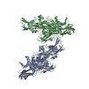
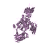
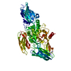
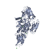
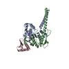
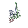
| Title | Role of Calcium Ions in the Activation and Activity of the Transglutaminase 3 Enzyme (3 calciums, active form) | ||||||
 Components Components | Protein-glutamine glutamyltransferase E | ||||||
 Keywords Keywords |  TRANSFERASE / Transglutaminase 3 / metalloenzyme / calcium ion TRANSFERASE / Transglutaminase 3 / metalloenzyme / calcium ion | ||||||
| Function / homology |  Function and homology informationprotein-glutamine gamma-glutamyltransferase / protein-glutamine gamma-glutamyltransferase activity / peptide cross-linking / hair follicle morphogenesis / acyltransferase activity / keratinization / catalytic activity / keratinocyte differentiation / extrinsic component of cytoplasmic side of plasma membrane / protein modification process ...protein-glutamine gamma-glutamyltransferase / protein-glutamine gamma-glutamyltransferase activity / peptide cross-linking / hair follicle morphogenesis / acyltransferase activity / keratinization / catalytic activity / keratinocyte differentiation / extrinsic component of cytoplasmic side of plasma membrane / protein modification process / calcium ion binding / structural molecule activity / protein-containing complex / extracellular exosome / cytoplasm Function and homology informationprotein-glutamine gamma-glutamyltransferase / protein-glutamine gamma-glutamyltransferase activity / peptide cross-linking / hair follicle morphogenesis / acyltransferase activity / keratinization / catalytic activity / keratinocyte differentiation / extrinsic component of cytoplasmic side of plasma membrane / protein modification process ...protein-glutamine gamma-glutamyltransferase / protein-glutamine gamma-glutamyltransferase activity / peptide cross-linking / hair follicle morphogenesis / acyltransferase activity / keratinization / catalytic activity / keratinocyte differentiation / extrinsic component of cytoplasmic side of plasma membrane / protein modification process / calcium ion binding / structural molecule activity / protein-containing complex / extracellular exosome / cytoplasmSimilarity search - Function | ||||||
| Biological species |  Homo sapiens (human) Homo sapiens (human) | ||||||
| Method | X-RAY DIFFRACTION / SYNCHROTRON / MOLECULAR REPLACEMENT / Resolution: 2.7 Å | ||||||
 Authors Authors | Ahvazi, B. | ||||||
 Citation Citation | Journal: J.Biol.Chem. / Year: 2003 Title: Roles of Calcium Ions in the Activation and Activity of the Transglutaminase 3 Enzyme Authors: Ahvazi, B. / Boeshans, K.M. / Idler, W. / Baxa, U. / Steinert, P.M. #1: Journal: Embo J. / Year: 2002Title: Three-dimensional structure of the human transglutaminase 3 enzyme:binding of calcium ions changes structure for activation Authors: Ahvazi, B. / Kim, H.C. / Kee, S.H. / Nemes, Z. / Steinert, P.M. #2: Journal: J.Struct.Biol. / Year: 2001Title: Crystallization and Preliminary X-ray Analysis of Human Transglutaminase 3 from Zymogen to Active Form Authors: Kim, H.C. / Nemes, Z. / Idler, W.W. / Hyde, C.C. / Steinert, P.M. / Ahvazi, B. | ||||||
| History |
| ||||||
| Remark 295 | NON-CRYSTALLOGRAPHIC SYMMETRY THE TRANSFORMATIONS PRESENTED ON THE MTRIX RECORDS BELOW DESCRIBE ... NON-CRYSTALLOGRAPHIC SYMMETRY THE TRANSFORMATIONS PRESENTED ON THE MTRIX RECORDS BELOW DESCRIBE NON-CRYSTALLOGRAPHIC RELATIONSHIPS AMONG ATOMS IN THIS ENTRY. APPLYING THE APPROPRIATE MTRIX TRANSFORMATION TO THE RESIDUES LISTED FIRST WILL YIELD APPROXIMATE COORDINATES FOR THE RESIDUES LISTED SECOND. CHAIN IDENTIFIERS GIVEN AS "?" REFER TO CHAINS FOR WHICH ATOMS ARE NOT FOUND IN THIS ENTRY. APPLIED TO TRANSFORMED TO TRANSFORM CHAIN RESIDUES CHAIN RESIDUES RMSD SSS M 1 A 1 .. 692 B 1 .. 692 0.408 WHERE SSS -> COLUMNS 8-10 OF MTRIX RECORDS REMARK: THE MATRIX TRANSFORMS CA ONLY. | ||||||
| Remark 400 | COMPOUND THE ENZYME WAS PROTEOLYZED WITH DISPASE I FOR ACTIVATION. | ||||||
| Remark 999 | SEQUENCE The following residues are noted as conflicts in the Swiss-Prot database: K562R, G654R ...SEQUENCE The following residues are noted as conflicts in the Swiss-Prot database: K562R, G654R (sequence database numbering). According to the author, residue 251 (sequence database numbering) is Asp and does not represent a mutation but a mistake in the Swiss-Prot database. |
- Structure visualization
| Structure viewer | Molecule: MolmilJmol/JSmol |
|---|
- Downloads & links
-Download
| PDBx/mmCIF format | 1nud.cif.gz | 276.4 KB | Display | PDBx/mmCIF format |
|---|---|---|---|---|
| PDB format | pdb1nud.ent.gz | 221.2 KB | Display | PDB format |
| PDBx/mmJSON format | 1nud.json.gz | Tree view | PDBx/mmJSON format | |
| Others |  Other downloads Other downloads |
-Validation report
| Arichive directory | https://data.pdbj.org/pub/pdb/validation_reports/nu/1nudftp://data.pdbj.org/pub/pdb/validation_reports/nu/1nud | HTTPS FTP |
|---|
-Related structure data
| Related structure data |  1nufC  1nugC  1l9nS S: Starting model for refinement C: citing same article ( |
|---|---|
| Similar structure data |

-Links
 PDBj
PDBj


- Assembly
| Deposited unit | 
| ||||||||
|---|---|---|---|---|---|---|---|---|---|
| 1 | 
| ||||||||
| 2 | 
| ||||||||
| Unit cell |
| ||||||||
| Noncrystallographic symmetry (NCS) | NCS oper: (Code: given Matrix: (1, 0.00023, -0.00133), Vector : |
-Components
| #1: Protein | Mass: 76670.500 Da / Num. of mol.: 2 / Mutation: F264L Source method: isolated from a genetically manipulated source Source: (gene. exp.) Homo sapiens (human) / Gene: TGM3 / Plasmid: Bac-N_Blue, Invitrogen / Cell line (production host): SF9 / Production host:   Spodoptera frugiperda (fall armyworm) Spodoptera frugiperda (fall armyworm)References: UniProt: Q08188, protein-glutamine gamma-glutamyltransferase#2: Chemical | ChemComp-CA /   Mass: 40.078 Da / Num. of mol.: 6 / Source method: obtained synthetically / Formula: Ca Mass: 40.078 Da / Num. of mol.: 6 / Source method: obtained synthetically / Formula: Ca#3: Chemical | ChemComp-CL / Chloride  Mass: 35.453 Da / Num. of mol.: 8 / Source method: obtained synthetically / Formula: Cl Mass: 35.453 Da / Num. of mol.: 8 / Source method: obtained synthetically / Formula: Cl#4: Chemical | ChemComp-BR / Bromide  Mass: 79.904 Da / Num. of mol.: 9 / Source method: obtained synthetically / Formula: Br Mass: 79.904 Da / Num. of mol.: 9 / Source method: obtained synthetically / Formula: Br#5: Water | ChemComp-HOH / | Water Mass: 18.015 Da / Num. of mol.: 398 / Source method: isolated from a natural source / Formula: H2O Mass: 18.015 Da / Num. of mol.: 398 / Source method: isolated from a natural source / Formula: H2O |
|---|
-Experimental details
-Experiment
| Experiment | Method: X-RAY DIFFRACTION / Number of used crystals: 1 |
|---|
- Sample preparation
| Crystal | Density Matthews: 2.62 Å3/Da / Density % sol: 53.12 % | ||||||||||||||||||||||||||||||||||||||||||||||||||||||||
|---|---|---|---|---|---|---|---|---|---|---|---|---|---|---|---|---|---|---|---|---|---|---|---|---|---|---|---|---|---|---|---|---|---|---|---|---|---|---|---|---|---|---|---|---|---|---|---|---|---|---|---|---|---|---|---|---|---|
| Crystal grow | Temperature: 288 K / Method: vapor diffusion, hanging drop / pH: 8.5 Details: 4% (w/v) Peg 20K, 100 mM Tris-HCl (pH 8.5) , VAPOR DIFFUSION, HANGING DROP, temperature 288K | ||||||||||||||||||||||||||||||||||||||||||||||||||||||||
| Crystal grow | *PLUS Temperature: 21 ℃ / pH: 8 | ||||||||||||||||||||||||||||||||||||||||||||||||||||||||
| Components of the solutions | *PLUS
|
-Data collection
| Diffraction |
| ||||||||||||||||||
|---|---|---|---|---|---|---|---|---|---|---|---|---|---|---|---|---|---|---|---|
| Diffraction source |
| ||||||||||||||||||
| Detector |
| ||||||||||||||||||
| Radiation | Monochromator: SI 111 channel / Protocol: SINGLE WAVELENGTH / Monochromatic (M) / Laue (L): M / Scattering type: x-ray | ||||||||||||||||||
| Radiation wavelength |
| ||||||||||||||||||
| Reflection | Resolution: 2.7→20 Å / Num. all: 41052 / Num. obs: 41052 / % possible obs: 93.7 % / Observed criterion σ(F): 2 / Observed criterion σ(I): 2 / Biso Wilson estimate: 23.8 Å2 / Net I/σ(I): 8.9 | ||||||||||||||||||
| Reflection shell | Resolution: 2.7→2.87 Å / % possible all: 88.4 | ||||||||||||||||||
| Reflection | *PLUS Lowest resolution: 20 Å / Num. measured all: 409995 | ||||||||||||||||||
| Reflection shell | *PLUS Lowest resolution: 2.8 Å / % possible obs: 88.4 % / Mean I/σ(I) obs: 2.5 |

- Processing
| Software |
| |||||||||||||||||||||||||||
|---|---|---|---|---|---|---|---|---|---|---|---|---|---|---|---|---|---|---|---|---|---|---|---|---|---|---|---|---|
| Refinement | Method to determine structure: MOLECULAR REPLACEMENT Starting model: PDB Entry 1L9N Resolution: 2.7→20 Å / Rfactor Rfree error: 0.004 / Isotropic thermal model: Isotropic / Cross valid method: THROUGHOUT / σ(F): 0 / Stereochemistry target values: Engh & Huber Details: NCS two fold averaging was employed during refinement
| |||||||||||||||||||||||||||
| Displacement parameters | Biso mean: 21.6 Å2
| |||||||||||||||||||||||||||
| Refine analyze |
| |||||||||||||||||||||||||||
| Refinement step | Cycle: LAST / Resolution: 2.7→20 Å
| |||||||||||||||||||||||||||
| Refine LS restraints |
| |||||||||||||||||||||||||||
| LS refinement shell | Resolution: 2.7→2.87 Å / Rfactor Rfree error: 0.015
| |||||||||||||||||||||||||||
| Refinement | *PLUS Lowest resolution: 20 Å / % reflection Rfree: 10 % | |||||||||||||||||||||||||||
| Solvent computation | *PLUS | |||||||||||||||||||||||||||
| Displacement parameters | *PLUS | |||||||||||||||||||||||||||
| Refine LS restraints | *PLUS
|
