 Movie
Movie Controller

[English] 日本語
 Yorodumi
- PDB-5h0n: Crystal structure of HIV-1 fusion inhibitor MT-WQ-IDL bound to gp... -
+ Open data

- Basic information
| Entry | Database: PDB / ID: 5h0n | ||||||
|---|---|---|---|---|---|---|---|
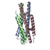
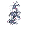
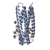
| Title | Crystal structure of HIV-1 fusion inhibitor MT-WQ-IDL bound to gp41 NHR | ||||||
 Components Components |
| ||||||
 Keywords Keywords |  VIRAL PROTEIN/VIRAL PROTEIN INHIBITOR / MT-WQ-IDL / HIV-1 / fusion inhibitor / VIRAL PROTEIN-VIRAL PROTEIN INHIBITOR complex VIRAL PROTEIN/VIRAL PROTEIN INHIBITOR / MT-WQ-IDL / HIV-1 / fusion inhibitor / VIRAL PROTEIN-VIRAL PROTEIN INHIBITOR complex | ||||||
| Function / homology |  Function and homology information Function and homology informationSynthesis and processing of ENV and VPU / evasion of host immune response / Alpha-defensins / Dectin-2 family / Binding and entry of HIV virion / positive regulation of plasma membrane raft polarization / positive regulation of receptor clustering / positive regulation of establishment of T cell polarity / virus-mediated perturbation of host defense response / host cell endosome membrane ...Synthesis and processing of ENV and VPU / evasion of host immune response / Alpha-defensins / Dectin-2 family / Binding and entry of HIV virion / positive regulation of plasma membrane raft polarization / positive regulation of receptor clustering / positive regulation of establishment of T cell polarity / virus-mediated perturbation of host defense response / host cell endosome membrane / actin filament organization / Assembly Of The HIV Virion / Budding and maturation of HIV virion / clathrin-dependent endocytosis of virus by host cell / viral protein processing / symbiont entry into host cell / fusion of virus membrane with host plasma membrane / fusion of virus membrane with host endosome membrane / viral envelope / virion attachment to host cell / host cell plasma membrane / virion membrane / structural molecule activity / membraneSimilarity search - Function | ||||||
| Biological species |   Human immunodeficiency virus 1 Human immunodeficiency virus 1 | ||||||
| Method | X-RAY DIFFRACTION / SYNCHROTRON / MOLECULAR REPLACEMENT / Resolution: 2.8 Å | ||||||
 Authors Authors | Zhu, Y. / Ye, S. / Zhang, R. | ||||||
 Citation Citation | Journal: J. Virol. / Year: 2017 Title: Creating an Artificial Tail Anchor as a Novel Strategy To Enhance the Potency of Peptide-Based HIV Fusion Inhibitors Authors: Su, S. / Zhu, Y. / Ye, S. / Qi, Q. / Xia, S. / Ma, Z. / Yu, F. / Wang, Q. / Zhang, R. / Jiang, S. / Lu, L. | ||||||
| History |
|
- Structure visualization
| Structure viewer | Molecule: MolmilJmol/JSmol |
|---|
- Downloads & links
-Download
| PDBx/mmCIF format | 5h0n.cif.gz | 99.3 KB | Display | PDBx/mmCIF format |
|---|---|---|---|---|
| PDB format | pdb5h0n.ent.gz | 77.8 KB | Display | PDB format |
| PDBx/mmJSON format | 5h0n.json.gz | Tree view | PDBx/mmJSON format | |
| Others |  Other downloads Other downloads |
-Validation report
| Arichive directory | https://data.pdbj.org/pub/pdb/validation_reports/h0/5h0nftp://data.pdbj.org/pub/pdb/validation_reports/h0/5h0n | HTTPS FTP |
|---|
-Related structure data
| Related structure data | 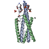 5cmzS S: Starting model for refinement |
|---|---|
| Similar structure data |


-Links
 PDBj
PDBj


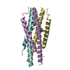
- Assembly
| Deposited unit | 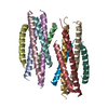
| ||||||||
|---|---|---|---|---|---|---|---|---|---|
| 1 | 
| ||||||||
| 2 | 
| ||||||||
| Unit cell |
|
-Components
| #1: Protein/peptide | Mass: 5252.146 Da / Num. of mol.: 6 / Source method: obtained synthetically / Source: (synth.) Human immunodeficiency virus 1 / References: UniProt: P04578*PLUS#2: Protein/peptide | Mass: 3931.463 Da / Num. of mol.: 6 / Source method: obtained synthetically / Source: (synth.) Human immunodeficiency virus 1#3: Water | ChemComp-HOH / | Water Mass: 18.015 Da / Num. of mol.: 7 / Source method: isolated from a natural source / Formula: H2O Mass: 18.015 Da / Num. of mol.: 7 / Source method: isolated from a natural source / Formula: H2O |
|---|
-Experimental details
-Experiment
| Experiment | Method: X-RAY DIFFRACTION / Number of used crystals: 1 |
|---|
- Sample preparation
| Crystal | Density Matthews: 2.63 Å3/Da / Density % sol: 53.31 % |
|---|---|
| Crystal grow | Temperature: 289 K / Method: vapor diffusion Details: 0.2 M Zinc Acetate, 0.1 M Imidazole: HCl pH 8.0, 20% (v/v) 1,4-Butanediol |
-Data collection
| Diffraction | Mean temperature: 100 K |
|---|---|
| Diffraction source | Source: SYNCHROTRON / Site: SSRF  / Beamline: BL19U1 / Wavelength: 0.97853 Å / Beamline: BL19U1 / Wavelength: 0.97853 Å |
| Detector | Type: DECTRIS PILATUS3 2M / Detector: PIXEL / Date: Apr 27, 2016 |
| Radiation | Protocol: SINGLE WAVELENGTH / Monochromatic (M) / Laue (L): M / Scattering type: x-ray |
| Radiation wavelength | Wavelength: 0.97853 Å / Relative weight: 1 |
| Reflection | Resolution: 2.8→39.6 Å / Num. obs: 12009 / % possible obs: 84.6 % / Redundancy: 2.5 % / Rmerge(I) obs: 0.079 / Net I/σ(I): 9.3 |
| Reflection shell | Resolution: 2.8→2.85 Å |
- Processing
| Software |
| ||||||||||||||||||||||||||||||||||||||||||
|---|---|---|---|---|---|---|---|---|---|---|---|---|---|---|---|---|---|---|---|---|---|---|---|---|---|---|---|---|---|---|---|---|---|---|---|---|---|---|---|---|---|---|---|
| Refinement | Method to determine structure: MOLECULAR REPLACEMENT Starting model: 5CMZ Resolution: 2.8→39.6 Å / SU ML: 0.41 / Cross valid method: FREE R-VALUE / σ(F): 1.38 / Phase error: 30.34
| ||||||||||||||||||||||||||||||||||||||||||
| Solvent computation | Shrinkage radii: 0.9 Å / VDW probe radii: 1.11 Å | ||||||||||||||||||||||||||||||||||||||||||
| Refinement step | Cycle: LAST / Resolution: 2.8→39.6 Å
| ||||||||||||||||||||||||||||||||||||||||||
| Refine LS restraints |
| ||||||||||||||||||||||||||||||||||||||||||
| LS refinement shell |
|
