 Movie
Movie Controller


+ Open data

- Basic information
| Entry | Database: PDB / ID: 5g4x | ||||||
|---|---|---|---|---|---|---|---|
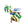
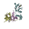
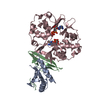
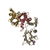
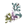
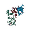
| Title | The crystal structure of the SHANK3 N-terminus | ||||||
 Components Components | SH3 AND MULTIPLE ANKYRIN REPEAT DOMAINS PROTEIN 3 | ||||||
 Keywords Keywords |  STRUCTURAL PROTEIN / SHANK3 / RAS / INTEGRIN. STRUCTURAL PROTEIN / SHANK3 / RAS / INTEGRIN. | ||||||
| Function / homology |  Function and homology information Function and homology informationresponse to interleukin-17 / regulation of AMPA glutamate receptor clustering / guanylate kinase-associated protein clustering / striatal medium spiny neuron differentiation / synaptic receptor adaptor activity / maintenance of postsynaptic density structure / RET signaling / positive regulation of glutamate receptor signaling pathway / postsynaptic density assembly / embryonic epithelial tube formation ...response to interleukin-17 / regulation of AMPA glutamate receptor clustering / guanylate kinase-associated protein clustering / striatal medium spiny neuron differentiation / synaptic receptor adaptor activity / maintenance of postsynaptic density structure / RET signaling / positive regulation of glutamate receptor signaling pathway / postsynaptic density assembly / embryonic epithelial tube formation / Neurexins and neuroligins / positive regulation of synapse structural plasticity / negative regulation of actin filament bundle assembly / vocal learning / negative regulation of cell volume / positive regulation of long-term neuronal synaptic plasticity / structural constituent of postsynaptic density / regulation of grooming behavior / NMDA glutamate receptor clustering / vocalization behavior / regulation of dendritic spine morphogenesis / regulation of behavioral fear response / neuron spine / AMPA glutamate receptor clustering / neural precursor cell proliferation / locomotion / dendritic spine morphogenesis / brain morphogenesis / regulation of long-term synaptic potentiation / positive regulation of AMPA receptor activity / long-term synaptic depression / regulation of postsynapse organization / ciliary membrane / exploration behavior / regulation of long-term synaptic depression / adult behavior / positive regulation of dendritic spine development / locomotory exploration behavior / postsynaptic density, intracellular component / social behavior / associative learning / positive regulation of excitatory postsynaptic potential / neuromuscular process controlling balance / glial cell proliferation / synapse assembly / ionotropic glutamate receptor binding / positive regulation of synaptic transmission, glutamatergic / locomotory behavior / learning / long-term synaptic potentiation / positive regulation of long-term synaptic potentiation / G protein-coupled receptor binding / regulation of synaptic plasticity / modulation of chemical synaptic transmission / memory / SH3 domain binding / : / MAPK cascade / gene expression / actin binding / scaffold protein binding / dendritic spine / postsynaptic density / learning or memory / neuron projection / glutamatergic synapse / protein-containing complex binding / zinc ion binding / identical protein binding / plasma membrane / cytoplasmSimilarity search - Function | ||||||
| Biological species |  RATTUS NORVEGICUS (Norway rat) RATTUS NORVEGICUS (Norway rat) | ||||||
| Method | X-RAY DIFFRACTION / SYNCHROTRON / MOLECULAR REPLACEMENT / Resolution: 2.166 Å | ||||||
 Authors Authors | Zacharchenko, T. / Barsukov, I. | ||||||
 Citation Citation | Journal: Nat. Cell Biol. / Year: 2017 Title: SHANK proteins limit integrin activation by directly interacting with Rap1 and R-Ras. Authors: Lilja, J. / Zacharchenko, T. / Georgiadou, M. / Jacquemet, G. / Franceschi, N. / Peuhu, E. / Hamidi, H. / Pouwels, J. / Martens, V. / Nia, F.H. / Beifuss, M. / Boeckers, T. / Kreienkamp, H.J. ...Authors: Lilja, J. / Zacharchenko, T. / Georgiadou, M. / Jacquemet, G. / Franceschi, N. / Peuhu, E. / Hamidi, H. / Pouwels, J. / Martens, V. / Nia, F.H. / Beifuss, M. / Boeckers, T. / Kreienkamp, H.J. / Barsukov, I.L. / Ivaska, J. | ||||||
| History |
|
- Structure visualization
| Structure viewer | Molecule: MolmilJmol/JSmol |
|---|
- Downloads & links
-Download
| PDBx/mmCIF format | 5g4x.cif.gz | 156.3 KB | Display | PDBx/mmCIF format |
|---|---|---|---|---|
| PDB format | pdb5g4x.ent.gz | 126.2 KB | Display | PDB format |
| PDBx/mmJSON format | 5g4x.json.gz | Tree view | PDBx/mmJSON format | |
| Others |  Other downloads Other downloads |
-Validation report
| Arichive directory | https://data.pdbj.org/pub/pdb/validation_reports/g4/5g4xftp://data.pdbj.org/pub/pdb/validation_reports/g4/5g4x | HTTPS FTP |
|---|
-Related structure data
| Related structure data | 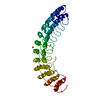 1n11S S: Starting model for refinement |
|---|---|
| Similar structure data |


-Links
 PDBj
PDBj
- Assembly
| Deposited unit | 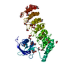
| ||||||||
|---|---|---|---|---|---|---|---|---|---|
| 1 |
| ||||||||
| Unit cell |
|
-Components
| #1: Protein | Mass: 38631.645 Da / Num. of mol.: 1 / Fragment: SPN-ARR, RESIDUES 1-348 / Source method: isolated from a natural source / Source: (natural) RATTUS NORVEGICUS (Norway rat) / References: UniProt: Q9JLU4 | ||
|---|---|---|---|
| #2: Chemical | ChemComp-EDO / Ethylene glycol  Mass: 62.068 Da / Num. of mol.: 30 / Source method: obtained synthetically / Formula: C2H6O2 Mass: 62.068 Da / Num. of mol.: 30 / Source method: obtained synthetically / Formula: C2H6O2#3: Water | ChemComp-HOH / | Water Mass: 18.015 Da / Num. of mol.: 258 / Source method: isolated from a natural source / Formula: H2O Mass: 18.015 Da / Num. of mol.: 258 / Source method: isolated from a natural source / Formula: H2O |
-Experimental details
-Experiment
| Experiment | Method: X-RAY DIFFRACTION / Number of used crystals: 1 |
|---|
- Sample preparation
| Crystal | Density Matthews: 3.81 Å3/Da / Density % sol: 67 % / Description: NONE |
|---|---|
| Crystal grow | pH: 7.5 / Details: 0.1M HEPES PH 7.5, 0.4M K/NA TARTRATE |
-Data collection
| Diffraction | Mean temperature: 100 K |
|---|---|
| Diffraction source | Source: SYNCHROTRON / Site: Diamond  / Beamline: I04-1 / Wavelength: 0.92 / Beamline: I04-1 / Wavelength: 0.92 |
| Detector | Type: DECTRIS PILATUS / Detector: PIXEL / Date: Dec 16, 2012 |
| Radiation | Protocol: SINGLE WAVELENGTH / Monochromatic (M) / Laue (L): M / Scattering type: x-ray |
| Radiation wavelength | Wavelength: 0.92 Å / Relative weight: 1 |
| Reflection | Resolution: 2.16→48.54 Å / Num. obs: 32995 / % possible obs: 100 % / Observed criterion σ(I): 4 / Redundancy: 12.2 % / Biso Wilson estimate: 34.17 Å2 / Rmerge(I) obs: 0.11 / Net I/σ(I): 14.7 |
| Reflection shell | Resolution: 2.17→2.3 Å / Redundancy: 12.6 % / Rmerge(I) obs: 0.6 / Mean I/σ(I) obs: 4.4 / % possible all: 100 |
- Processing
| Software |
| |||||||||||||||||||||||||||||||||||||||||||||||||||||||||||||||||||||||||||||||||||||||||||
|---|---|---|---|---|---|---|---|---|---|---|---|---|---|---|---|---|---|---|---|---|---|---|---|---|---|---|---|---|---|---|---|---|---|---|---|---|---|---|---|---|---|---|---|---|---|---|---|---|---|---|---|---|---|---|---|---|---|---|---|---|---|---|---|---|---|---|---|---|---|---|---|---|---|---|---|---|---|---|---|---|---|---|---|---|---|---|---|---|---|---|---|---|
| Refinement | Method to determine structure: MOLECULAR REPLACEMENT Starting model: PDB ENTRY 1N11 Resolution: 2.166→48.538 Å / SU ML: 0.18 / σ(F): 1.34 / Phase error: 16.6 / Stereochemistry target values: ML
| |||||||||||||||||||||||||||||||||||||||||||||||||||||||||||||||||||||||||||||||||||||||||||
| Solvent computation | Shrinkage radii: 0.9 Å / VDW probe radii: 1.11 Å / Solvent model: FLAT BULK SOLVENT MODEL | |||||||||||||||||||||||||||||||||||||||||||||||||||||||||||||||||||||||||||||||||||||||||||
| Refinement step | Cycle: LAST / Resolution: 2.166→48.538 Å
| |||||||||||||||||||||||||||||||||||||||||||||||||||||||||||||||||||||||||||||||||||||||||||
| Refine LS restraints |
| |||||||||||||||||||||||||||||||||||||||||||||||||||||||||||||||||||||||||||||||||||||||||||
| LS refinement shell |
| |||||||||||||||||||||||||||||||||||||||||||||||||||||||||||||||||||||||||||||||||||||||||||
| Refinement TLS params. | Method: refined / Refine-ID: X-RAY DIFFRACTION
| |||||||||||||||||||||||||||||||||||||||||||||||||||||||||||||||||||||||||||||||||||||||||||
| Refinement TLS group |
|
