[English] 日本語
Yorodumi
- PDB-5fs4: Bacteriophage AP205 coat protein -

+
Open data


ID or keywords:

Loading...

-
Basic information

Entry
Database: PDB / ID: 5fs4
TitleBacteriophage AP205 coat protein
ComponentsAP205 BACTERIOPHAGE COAT PROTEIN
KeywordsVIRAL PROTEIN / SMALL RNA PHAGE / COAT PROTEIN / AP205
Function / homologyviral capsid / Coat protein
Function and homology information
Biological speciesACINETOBACTER PHAGE AP205 (virus)
MethodX-RAY DIFFRACTION / SYNCHROTRON / MAD / Resolution: 1.73 Å
AuthorsShishovs, M. / Tars, K.
Citation
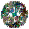
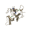
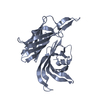
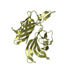
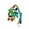
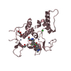
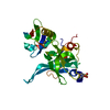
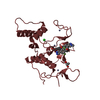
Journal: J Mol Biol / Year: 2016
Title: Structure of AP205 Coat Protein Reveals Circular Permutation in ssRNA Bacteriophages.
Authors: Mihails Shishovs / Janis Rumnieks / Christoph Diebolder / Kristaps Jaudzems / Loren B Andreas / Jan Stanek / Andris Kazaks / Svetlana Kotelovica / Inara Akopjana / Guido Pintacuda / Roman I ...Authors: Mihails Shishovs / Janis Rumnieks / Christoph Diebolder / Kristaps Jaudzems / Loren B Andreas / Jan Stanek / Andris Kazaks / Svetlana Kotelovica / Inara Akopjana / Guido Pintacuda / Roman I Koning / Kaspars Tars /
Abstract: AP205 is a single-stranded RNA bacteriophage that has a coat protein sequence not similar to any other known single-stranded RNA phage. Here, we report an atomic-resolution model of the AP205 virus- ...AP205 is a single-stranded RNA bacteriophage that has a coat protein sequence not similar to any other known single-stranded RNA phage. Here, we report an atomic-resolution model of the AP205 virus-like particle based on a crystal structure of an unassembled coat protein dimer and a cryo-electron microscopy reconstruction of the assembled particle, together with secondary structure information from site-specific solid-state NMR data. The AP205 coat protein dimer adopts the conserved Leviviridae coat protein fold except for the N-terminal region, which forms a beta-hairpin in the other known single-stranded RNA phages. AP205 has a similar structure at the same location formed by N- and C-terminal beta-strands, making it a circular permutant compared to the other coat proteins. The permutation moves the coat protein termini to the most surface-exposed part of the assembled particle, which explains its increased tolerance to long N- and C-terminal fusions.
History
DepositionDec 29, 2015Deposition site: PDBE / Processing site: PDBE
Revision 1.0Sep 21, 2016Provider: repository / Type: Initial release
Revision 1.1Nov 9, 2016Group: Database references
Revision 1.2Jan 17, 2018Group: Data collection / Category: diffrn_source / Item: _diffrn_source.pdbx_synchrotron_site
Revision 1.3Jan 30, 2019Group: Data collection / Experimental preparation
Category: database_PDB_rev / database_PDB_rev_record ...database_PDB_rev / database_PDB_rev_record / exptl_crystal_grow / struct_biol
Item: _exptl_crystal_grow.method / _exptl_crystal_grow.temp

-
Structure visualization

Structure viewerMolecule:
MolmilJmol/JSmol

Downloads & links

-
Assembly

Deposited unit
A: AP205 BACTERIOPHAGE COAT PROTEIN
B: AP205 BACTERIOPHAGE COAT PROTEIN


Theoretical massNumber of molelcules
Total (without water)28,1502
Polymers28,1502
Non-polymers00
Water3,747208
1


  • Idetical with deposited unit
  • defined by author&software
TypeNameSymmetry operationNumber
identity operation1_555x,y,z1
Buried area7180 Å2
ΔGint-46.3 kcal/mol
Surface area12020 Å2
MethodPISA
Unit cell
Length a, b, c (Å)28.600, 114.930, 42.800
Angle α, β, γ (deg.)90.00, 98.14, 90.00
Int Tables number4
Space group name H-MP1211
Noncrystallographic symmetry (NCS)NCS oper: (Code: given
Matrix: (0.8754, 0.4026, 0.2677), (0.4028, -0.9135, 0.05679), (0.2674, 0.05811, -0.9618)
Vector: -23.01, 62.91, 66.86)

-
Components

#1: Protein AP205 BACTERIOPHAGE COAT PROTEIN


Mass: 14074.790 Da / Num. of mol.: 2 / Mutation: YES
Source method: isolated from a genetically manipulated source
Source: (gene. exp.) ACINETOBACTER PHAGE AP205 (virus) / Plasmid: PRSF-TEVDUET / Production host: ESCHERICHIA COLI (E. coli) / Strain (production host): BL21(DE3) / References: UniProt: Q9AZ42
#2: Water ChemComp-HOH / water / Water


Mass: 18.015 Da / Num. of mol.: 208 / Source method: isolated from a natural source / Formula: H2O
Sequence detailsN-TERMINAL SERINE AS A RESULT OF RECOMBINANT CONSTRUCT INSERTION IN A PLASMID

-
Experimental details

-
Experiment

ExperimentMethod: X-RAY DIFFRACTION / Number of used crystals: 2

-
Sample preparation

CrystalDensity Matthews: 2.49 Å3/Da / Density % sol: 50.57 % / Description: NONE
Crystal growTemperature: 294 K / Method: vapor diffusion, sitting drop / pH: 8
Details: 20 % ETHYLENE GLYCOL AQUEOUS SOLUTION, 7 MG/ML PROTEIN IN 40 MM TRIS-HCL PH 8,0, 300 MM NACL, VAPOR DIFFUSION SITTING DROP METHOR, 0.7 MIKROL PRECIPITANT PLUS 1 MIKROL PROTEIN,TEMPERATURE 294 K

-
Data collection

DiffractionMean temperature: 100 K
Diffraction sourceSource: SYNCHROTRON / Site: MAX II / Beamline: I911-3 / Wavelength: 1
DetectorType: MARRESEARCH / Detector: CCD / Date: Dec 8, 2015 / Details: MULTILAYER
RadiationMonochromator: SI CRYSTAL / Protocol: MAD / Monochromatic (M) / Laue (L): M / Scattering type: x-ray
Radiation wavelengthWavelength: 1 Å / Relative weight: 1
ReflectionResolution: 1.73→57.45 Å / Num. obs: 28660 / % possible obs: 99.8 % / Observed criterion σ(I): 3 / Redundancy: 3.2 % / Biso Wilson estimate: 5 Å2 / Rmerge(I) obs: 0.17 / Net I/σ(I): 4.6
Reflection shellResolution: 1.73→1.82 Å / Redundancy: 2.8 % / Rmerge(I) obs: 0.63 / Mean I/σ(I) obs: 1.6 / % possible all: 99.1

-
Processing

Software
NameVersionClassification
REFMAC5.8.0103refinement
MOSFLMdata reduction
SCALAdata scaling
BUCCANEERphasing
RefinementMethod to determine structure: MAD
Starting model: NONE

Resolution: 1.73→57.53 Å / Cor.coef. Fo:Fc: 0.891 / Cor.coef. Fo:Fc free: 0.822 / SU B: 4.263 / SU ML: 0.134 / Cross valid method: THROUGHOUT / ESU R: 0.133 / ESU R Free: 0.132 / Stereochemistry target values: MAXIMUM LIKELIHOOD
Details: HYDROGENS HAVE BEEN ADDED IN THE RIDING POSITIONS. HYDROGENS HAVE BEEN ADDED IN THE RIDING POSITIONS U VALUES REFINED INDIVIDUALLY
RfactorNum. reflection% reflectionSelection details
Rfree0.27454 1416 4.9 %RANDOM
Rwork0.23026 ---
obs0.23246 27197 99.76 %-
Solvent computationIon probe radii: 0.8 Å / Shrinkage radii: 0.8 Å / VDW probe radii: 1.2 Å / Solvent model: MASK
Displacement parametersBiso mean: 14.587 Å2
Baniso -1Baniso -2Baniso -3
1-1.87 Å20 Å20.17 Å2
2---0.8 Å20 Å2
3----1.08 Å2
Refinement stepCycle: LAST / Resolution: 1.73→57.53 Å
ProteinNucleic acidLigandSolventTotal
Num. atoms1836 0 0 208 2044
Refine LS restraints
Refine-IDTypeDev idealDev ideal targetNumber
X-RAY DIFFRACTIONr_bond_refined_d0.0170.021866
X-RAY DIFFRACTIONr_bond_other_d0.0020.021780
X-RAY DIFFRACTIONr_angle_refined_deg1.8321.9552537
X-RAY DIFFRACTIONr_angle_other_deg0.98234094
X-RAY DIFFRACTIONr_dihedral_angle_1_deg6.9075240
X-RAY DIFFRACTIONr_dihedral_angle_2_deg38.64524.52173
X-RAY DIFFRACTIONr_dihedral_angle_3_deg11.96515305
X-RAY DIFFRACTIONr_dihedral_angle_4_deg17.1071512
X-RAY DIFFRACTIONr_chiral_restr0.1080.2301
X-RAY DIFFRACTIONr_gen_planes_refined0.0090.0212113
X-RAY DIFFRACTIONr_gen_planes_other0.0010.02403
X-RAY DIFFRACTIONr_nbd_refined
X-RAY DIFFRACTIONr_nbd_other
X-RAY DIFFRACTIONr_nbtor_refined
X-RAY DIFFRACTIONr_nbtor_other
X-RAY DIFFRACTIONr_xyhbond_nbd_refined
X-RAY DIFFRACTIONr_xyhbond_nbd_other
X-RAY DIFFRACTIONr_metal_ion_refined
X-RAY DIFFRACTIONr_metal_ion_other
X-RAY DIFFRACTIONr_symmetry_vdw_refined
X-RAY DIFFRACTIONr_symmetry_vdw_other
X-RAY DIFFRACTIONr_symmetry_hbond_refined
X-RAY DIFFRACTIONr_symmetry_hbond_other
X-RAY DIFFRACTIONr_symmetry_metal_ion_refined
X-RAY DIFFRACTIONr_symmetry_metal_ion_other
X-RAY DIFFRACTIONr_mcbond_it1.4081.343975
X-RAY DIFFRACTIONr_mcbond_other1.3521.34974
X-RAY DIFFRACTIONr_mcangle_it2.2721.9931210
X-RAY DIFFRACTIONr_mcangle_other
X-RAY DIFFRACTIONr_scbond_it1.5281.474891
X-RAY DIFFRACTIONr_scbond_other
X-RAY DIFFRACTIONr_scangle_it
X-RAY DIFFRACTIONr_scangle_other
X-RAY DIFFRACTIONr_long_range_B_refined
X-RAY DIFFRACTIONr_long_range_B_other
X-RAY DIFFRACTIONr_rigid_bond_restr
X-RAY DIFFRACTIONr_sphericity_free
X-RAY DIFFRACTIONr_sphericity_bonded
LS refinement shellResolution: 1.726→1.771 Å / Total num. of bins used: 20
RfactorNum. reflection% reflection
Rfree0.318 92 -
Rwork0.27 2009 -
obs--98.27 %

+
About Yorodumi

-
News

-
Feb 9, 2022. New format data for meta-information of EMDB entries

New format data for meta-information of EMDB entries

  • Version 3 of the EMDB header file is now the official format.
  • The previous official version 1.9 will be removed from the archive.

Related info.:EMDB header

External links:wwPDB to switch to version 3 of the EMDB data model

-
Aug 12, 2020. Covid-19 info

Covid-19 info

URL: https://pdbj.org/emnavi/covid19.php

New page: Covid-19 featured information page in EM Navigator.

Related info.:Covid-19 info / Mar 5, 2020. Novel coronavirus structure data

+
Mar 5, 2020. Novel coronavirus structure data

Novel coronavirus structure data

Related info.:Yorodumi Speices / Aug 12, 2020. Covid-19 info

External links:COVID-19 featured content - PDBj / Molecule of the Month (242):Coronavirus Proteases

+
Jan 31, 2019. EMDB accession codes are about to change! (news from PDBe EMDB page)

EMDB accession codes are about to change! (news from PDBe EMDB page)

  • The allocation of 4 digits for EMDB accession codes will soon come to an end. Whilst these codes will remain in use, new EMDB accession codes will include an additional digit and will expand incrementally as the available range of codes is exhausted. The current 4-digit format prefixed with “EMD-” (i.e. EMD-XXXX) will advance to a 5-digit format (i.e. EMD-XXXXX), and so on. It is currently estimated that the 4-digit codes will be depleted around Spring 2019, at which point the 5-digit format will come into force.
  • The EM Navigator/Yorodumi systems omit the EMD- prefix.

Related info.:Q: What is EMD? / ID/Accession-code notation in Yorodumi/EM Navigator

External links:EMDB Accession Codes are Changing Soon! / Contact to PDBj

+
Jul 12, 2017. Major update of PDB

Major update of PDB

  • wwPDB released updated PDB data conforming to the new PDBx/mmCIF dictionary.
  • This is a major update changing the version number from 4 to 5, and with Remediation, in which all the entries are updated.
  • In this update, many items about electron microscopy experimental information are reorganized (e.g. em_software).
  • Now, EM Navigator and Yorodumi are based on the updated data.

External links:wwPDB Remediation / Enriched Model Files Conforming to OneDep Data Standards Now Available in the PDB FTP Archive

-
Yorodumi

Thousand views of thousand structures

  • Yorodumi is a browser for structure data from EMDB, PDB, SASBDB, etc.
  • This page is also the successor to EM Navigator detail page, and also detail information page/front-end page for Omokage search.
  • The word "yorodu" (or yorozu) is an old Japanese word meaning "ten thousand". "mi" (miru) is to see.

Related info.:EMDB / PDB / SASBDB / Comparison of 3 databanks / Yorodumi Search / Aug 31, 2016. New EM Navigator & Yorodumi / Yorodumi Papers / Jmol/JSmol / Function and homology information / Changes in new EM Navigator and Yorodumi

Read more