 Movie
Movie Controller

[English] 日本語
 Yorodumi
- PDB-1ij6: CA2+-BOUND STRUCTURE OF MULTIDOMAIN EF-HAND PROTEIN, CBP40, FROM ... -
+ Open data

- Basic information
| Entry | Database: PDB / ID: 1ij6 | ||||||
|---|---|---|---|---|---|---|---|
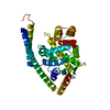
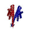
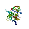
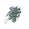
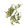
| Title | CA2+-BOUND STRUCTURE OF MULTIDOMAIN EF-HAND PROTEIN, CBP40, FROM TRUE SLIME MOLD | ||||||
 Components Components | PLASMODIAL SPECIFIC LAV1-2 PROTEIN | ||||||
 Keywords Keywords | METAL BINDING PROTEIN / FOURTY KDA CALCIUM BINDING PROTEIN / CBP40 | ||||||
| Function / homology |  Function and homology information Function and homology information | ||||||
| Biological species |   Physarum polycephalum (eukaryote) Physarum polycephalum (eukaryote) | ||||||
| Method | X-RAY DIFFRACTION / SYNCHROTRON / MOLECULAR REPLACEMENT / Resolution: 3.1 Å | ||||||
 Authors Authors | Iwasaki, W. / Sasaki, H. / Nakamura, A. / Kohama, K. / Tanokura, M. | ||||||
 Citation Citation | Journal: Structure / Year: 2003 Title: Metal-Free and Ca(2+)-Bound Structures of a Multidomain EF-Hand Protein, CBP40, from the Lower Eukaryote Physarum polycephalum Authors: Iwasaki, W. / Sasaki, H. / Nakamura, A. / Kohama, K. / Tanokura, M. | ||||||
| History |
|
- Structure visualization
| Structure viewer | Molecule: MolmilJmol/JSmol |
|---|
- Downloads & links
-Download
| PDBx/mmCIF format | 1ij6.cif.gz | 73.4 KB | Display | PDBx/mmCIF format |
|---|---|---|---|---|
| PDB format | pdb1ij6.ent.gz | 54.5 KB | Display | PDB format |
| PDBx/mmJSON format | 1ij6.json.gz | Tree view | PDBx/mmJSON format | |
| Others |  Other downloads Other downloads |
-Validation report
| Arichive directory | https://data.pdbj.org/pub/pdb/validation_reports/ij/1ij6ftp://data.pdbj.org/pub/pdb/validation_reports/ij/1ij6 | HTTPS FTP |
|---|
-Related structure data


-Links
 PDBj
PDBj- Assembly
| Deposited unit | 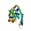
| ||||||||
|---|---|---|---|---|---|---|---|---|---|
| 1 |
| ||||||||
| Unit cell |
|
-Components
| #1: Protein | Mass: 37137.617 Da / Num. of mol.: 1 / Fragment: RESIDUES 33-355 Source method: isolated from a genetically manipulated source Source: (gene. exp.) Physarum polycephalum (eukaryote) / Plasmid: PET21D / Production host:  Escherichia coli (E. coli) / References: UniProt: P14725 Escherichia coli (E. coli) / References: UniProt: P14725 |
|---|---|
| #2: Chemical | ChemComp-CA /   Mass: 40.078 Da / Num. of mol.: 4 / Source method: obtained synthetically / Formula: Ca Mass: 40.078 Da / Num. of mol.: 4 / Source method: obtained synthetically / Formula: Ca |
-Experimental details
-Experiment
| Experiment | Method: X-RAY DIFFRACTION / Number of used crystals: 2 |
|---|
- Sample preparation
| Crystal | Density Matthews: 3.37 Å3/Da / Density % sol: 63.55 % | ||||||||||||||||||||||||||||||||||||||||||||||||||||||||
|---|---|---|---|---|---|---|---|---|---|---|---|---|---|---|---|---|---|---|---|---|---|---|---|---|---|---|---|---|---|---|---|---|---|---|---|---|---|---|---|---|---|---|---|---|---|---|---|---|---|---|---|---|---|---|---|---|---|
| Crystal grow | Temperature: 298 K / Method: vapor diffusion, hanging drop / pH: 7.6 Details: Ammonium Sulfate, Calcium chloride, pH 7.6, VAPOR DIFFUSION, HANGING DROP, temperature 298K | ||||||||||||||||||||||||||||||||||||||||||||||||||||||||
| Crystal grow | *PLUS Temperature: 25 ℃ / Details: Iwasaki, W., (1999) J.Biochem., 126, 7. | ||||||||||||||||||||||||||||||||||||||||||||||||||||||||
| Components of the solutions | *PLUS
|
-Data collection
| Diffraction | Mean temperature: 298 K |
|---|---|
| Diffraction source | Source: SYNCHROTRON / Site: Photon Factory  / Beamline: BL-6A / Wavelength: 1 Å / Beamline: BL-6A / Wavelength: 1 Å |
| Detector | Type: WEISSENBERG / Detector: DIFFRACTOMETER / Date: Dec 22, 1997 / Details: focusing mirrors |
| Radiation | Monochromator: Si(111) / Protocol: SINGLE WAVELENGTH / Monochromatic (M) / Laue (L): M / Scattering type: x-ray |
| Radiation wavelength | Wavelength: 1 Å / Relative weight: 1 |
| Reflection | Resolution: 3.1→38.1 Å / Num. all: 13647 / Num. obs: 34014 / % possible obs: 97.1 % / Observed criterion σ(I): 2 / Redundancy: 2.5 % / Rmerge(I) obs: 0.097 |
| Reflection shell | Resolution: 3.12→3.23 Å / Rmerge(I) obs: 0.342 / % possible all: 99.6 |
| Reflection | *PLUS Num. obs: 13647 / Num. measured all: 34014 |
- Processing
| Software |
| ||||||||||||||||||||
|---|---|---|---|---|---|---|---|---|---|---|---|---|---|---|---|---|---|---|---|---|---|
| Refinement | Method to determine structure: MOLECULAR REPLACEMENT / Resolution: 3.1→10 Å / σ(F): 0 / σ(I): 2 / Stereochemistry target values: Engh & Huber
| ||||||||||||||||||||
| Refinement step | Cycle: LAST / Resolution: 3.1→10 Å
| ||||||||||||||||||||
| Refine LS restraints |
| ||||||||||||||||||||
| Refinement | *PLUS % reflection Rfree: 5 % | ||||||||||||||||||||
| Solvent computation | *PLUS | ||||||||||||||||||||
| Displacement parameters | *PLUS | ||||||||||||||||||||
| Refine LS restraints | *PLUS Type: c_angle_deg / Dev ideal: 1.38 |
