 Movie
Movie Controller

[English] 日本語
 Yorodumi
- PDB-5f3o: Crystal structure of EhRNaseIII229 from Entamoeba histolytica com... -
+ Open data

- Basic information
| Entry | Database: PDB / ID: 5f3o | ||||||
|---|---|---|---|---|---|---|---|
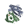
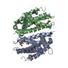
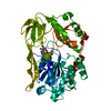
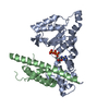
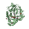
| Title | Crystal structure of EhRNaseIII229 from Entamoeba histolytica complexed with Mn2+ | ||||||
 Components Components | Putative uncharacterized protein | ||||||
 Keywords Keywords |  RNA BINDING PROTEIN / RNaseIII / non canonical Dicer / RNA processing / Entamoeba histolytica RNA BINDING PROTEIN / RNaseIII / non canonical Dicer / RNA processing / Entamoeba histolytica | ||||||
| Function / homology | ribonuclease III activity / Ribonuclease III, endonuclease domain superfamily / RNA processing / metal ion binding / : / Uncharacterized protein Function and homology information Function and homology information | ||||||
| Biological species |   Entamoeba histolytica (eukaryote) Entamoeba histolytica (eukaryote) | ||||||
| Method | X-RAY DIFFRACTION / SYNCHROTRON / Resolution: 2.05 Å | ||||||
 Authors Authors | Yu, X. / Gan, J.H. / Ma, J.B. | ||||||
 Citation Citation | Journal: To Be Published Title: Structural and functional study of a noncanonical Dicer protein in Entamoeba histolytica Authors: Yu, X. / Gan, J.H. / Ma, J.B. | ||||||
| History |
|
- Structure visualization
| Structure viewer | Molecule: MolmilJmol/JSmol |
|---|
- Downloads & links
-Download
| PDBx/mmCIF format | 5f3o.cif.gz | 91.8 KB | Display | PDBx/mmCIF format |
|---|---|---|---|---|
| PDB format | pdb5f3o.ent.gz | 69.7 KB | Display | PDB format |
| PDBx/mmJSON format | 5f3o.json.gz | Tree view | PDBx/mmJSON format | |
| Others |  Other downloads Other downloads |
-Validation report
| Arichive directory | https://data.pdbj.org/pub/pdb/validation_reports/f3/5f3oftp://data.pdbj.org/pub/pdb/validation_reports/f3/5f3o | HTTPS FTP |
|---|
-Related structure data


-Links
 PDBj
PDBj

- Assembly
| Deposited unit | 
| ||||||||
|---|---|---|---|---|---|---|---|---|---|
| 1 |
| ||||||||
| Unit cell |
|
-Components
| #1: Protein | Mass: 22893.994 Da / Num. of mol.: 2 / Fragment: UNP RESIDUES 2-195 Source method: isolated from a genetically manipulated source Source: (gene. exp.) Entamoeba histolytica (eukaryote) / Gene: EHI_068740 / Production host:  Escherichia coli (E. coli) / References: UniProt: C4LU64 Escherichia coli (E. coli) / References: UniProt: C4LU64#2: Chemical | Sulfate  Mass: 96.063 Da / Num. of mol.: 2 / Source method: obtained synthetically / Formula: SO4 Mass: 96.063 Da / Num. of mol.: 2 / Source method: obtained synthetically / Formula: SO4#3: Chemical |   Mass: 54.938 Da / Num. of mol.: 2 / Source method: obtained synthetically / Formula: Mn Mass: 54.938 Da / Num. of mol.: 2 / Source method: obtained synthetically / Formula: Mn#4: Water | ChemComp-HOH / | Water Mass: 18.015 Da / Num. of mol.: 100 / Source method: isolated from a natural source / Formula: H2O Mass: 18.015 Da / Num. of mol.: 100 / Source method: isolated from a natural source / Formula: H2O |
|---|
-Experimental details
-Experiment
| Experiment | Method: X-RAY DIFFRACTION |
|---|
- Sample preparation
| Crystal | Density Matthews: 2.29 Å3/Da / Density % sol: 46.18 % |
|---|---|
| Crystal grow | Temperature: 289 K / Method: vapor diffusion, sitting drop / pH: 7.5 / Details: 1.6M (NH4)2SO4, 0.01M MgCl2, 0.05M Tris-HCl pH 7.5 |
-Data collection
| Diffraction | Mean temperature: 100 K |
|---|---|
| Diffraction source | Source: SYNCHROTRON / Site: SSRF  / Beamline: BL17U / Wavelength: 1 Å / Beamline: BL17U / Wavelength: 1 Å |
| Detector | Type: ADSC QUANTUM 315 / Detector: CCD / Date: Apr 24, 2014 |
| Radiation | Protocol: SINGLE WAVELENGTH / Monochromatic (M) / Laue (L): M / Scattering type: x-ray |
| Radiation wavelength | Wavelength: 1 Å / Relative weight: 1 |
| Reflection | Resolution: 2.05→50 Å / Num. obs: 25785 / % possible obs: 95.3 % / Redundancy: 5.5 % / Net I/σ(I): 16.4 |
- Processing
| Software |
| ||||||||||||||||||||||||||||||||||||||||||||||||||||||||||||||||||||||
|---|---|---|---|---|---|---|---|---|---|---|---|---|---|---|---|---|---|---|---|---|---|---|---|---|---|---|---|---|---|---|---|---|---|---|---|---|---|---|---|---|---|---|---|---|---|---|---|---|---|---|---|---|---|---|---|---|---|---|---|---|---|---|---|---|---|---|---|---|---|---|---|
| Refinement | Resolution: 2.05→44.864 Å / SU ML: 0.25 / Cross valid method: THROUGHOUT / σ(F): 1.39 / Phase error: 23.24 / Stereochemistry target values: ML
| ||||||||||||||||||||||||||||||||||||||||||||||||||||||||||||||||||||||
| Solvent computation | Shrinkage radii: 0.9 Å / VDW probe radii: 1.11 Å / Solvent model: FLAT BULK SOLVENT MODEL | ||||||||||||||||||||||||||||||||||||||||||||||||||||||||||||||||||||||
| Refinement step | Cycle: LAST / Resolution: 2.05→44.864 Å
| ||||||||||||||||||||||||||||||||||||||||||||||||||||||||||||||||||||||
| Refine LS restraints |
| ||||||||||||||||||||||||||||||||||||||||||||||||||||||||||||||||||||||
| LS refinement shell |
|
