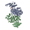
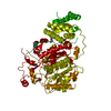
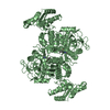
Mass: 69044.039 Da / Num. of mol.: 2 Source method: isolated from a genetically manipulated source Source: (gene. exp.) Bacillus subtilis (strain 168) (bacteria) Strain: 168 / Gene: pksL, outG, pksA, pksX, BSU17190 / Production host: Escherichia coli (E. coli) / References: UniProt: Q05470
-
Experimental details
-
Experiment
Experiment
Method: X-RAY DIFFRACTION
-
Sample preparation
Crystal
Density Matthews: 2.92 Å3/Da / Density % sol: 57.94 %
Crystal grow
Temperature: 298 K / Method: vapor diffusion, sitting drop Details: 150 mM (NH4)2SO4, 15% PEG 4000 (v/v), 100 mM MES pH 6.0 mixed with 15 mg/ml protein in 150 NaCl and 20 mM HEPES pH 7.5 in 1:2 ratio
-
Data collection
Diffraction
Mean temperature: 100 K
Diffraction source
Source: SYNCHROTRON / Site: ALS / Beamline: 5.0.3 / Wavelength: 0.977 Å
Protocol: SINGLE WAVELENGTH / Monochromatic (M) / Laue (L): M / Scattering type: x-ray
Radiation wavelength
Wavelength: 0.977 Å / Relative weight: 1
Reflection
Resolution: 3.1→39.73 Å / Num. obs: 24128 / % possible obs: 94.1 % / Redundancy: 1.9 % / Net I/σ(I): 2.81
-
Processing
Software
Name
Version
Classification
REFMAC
5.8.0107
refinement
HKL-2000
datareduction
HKL-2000
datascaling
PHASER
phasing
Refinement
Resolution: 3.1→39.73 Å / Cor.coef. Fo:Fc: 0.922 / Cor.coef. Fo:Fc free: 0.881 / Cross valid method: THROUGHOUT / ESU R Free: 0.519 / Stereochemistry target values: MAXIMUM LIKELIHOOD / Details: HYDROGENS HAVE BEEN ADDED IN THE RIDING POSITIONS
Rfactor
Num. reflection
% reflection
Selection details
Rfree
0.26841
1285
5.1 %
RANDOM
Rwork
0.2186
-
-
-
obs
0.22121
24128
94.08 %
-
Solvent computation
Ion probe radii: 0.8 Å / Shrinkage radii: 0.8 Å / VDW probe radii: 1.2 Å / Solvent model: MASK
 Movie
Movie Controller

 Yorodumi
 Open data
 Basic information
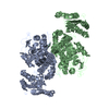
 Components
 Keywords
 HYDROLASE / trans-AT /
  Function and homology information
 Authors
 United States, 1items
  Citation
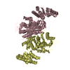
 Structure visualization
 Downloads & links
 Other downloads


 PDBj
PDBj

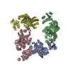
 Assembly
 Sample preparation
 Processing