 Movie
Movie Controller

[English] 日本語
 Yorodumi
- PDB-4na2: Crystal Structure of the second ketosynthase from the bacillaene ... -
+ Open data

- Basic information
| Entry | Database: PDB / ID: 4na2 | ||||||
|---|---|---|---|---|---|---|---|
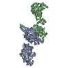
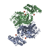
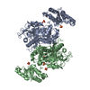
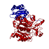
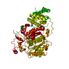
| Title | Crystal Structure of the second ketosynthase from the bacillaene polyketide synthase bound to its natural intermediate | ||||||
 Components Components | Polyketide synthase PksJ | ||||||
 Keywords Keywords |  TRANSFERASE / condensing enzyme fold TRANSFERASE / condensing enzyme fold | ||||||
| Function / homology |  Function and homology information Function and homology informationDIM/DIP cell wall layer assembly / organonitrogen compound biosynthetic process / fatty acid synthase activity / phosphopantetheine binding / ligase activity / 3-oxoacyl-[acyl-carrier-protein] synthase activity / antibiotic biosynthetic process / fatty acid biosynthetic process / cytoplasmSimilarity search - Function | ||||||
| Biological species |  Bacillus subtilis subsp. subtilis (bacteria) Bacillus subtilis subsp. subtilis (bacteria) | ||||||
| Method | X-RAY DIFFRACTION / SYNCHROTRON / MOLECULAR REPLACEMENT / Resolution: 2.3 Å | ||||||
 Authors Authors | Gay, D.C. / Gay, G.R. / Keatinge-Clay, A.T. | ||||||
 Citation Citation | Journal: Structure / Year: 2014 Title: A close look at a ketosynthase from a trans-acyltransferase modular polyketide synthase. Authors: Gay, D.C. / Gay, G. / Axelrod, A.J. / Jenner, M. / Kohlhaas, C. / Kampa, A. / Oldham, N.J. / Piel, J. / Keatinge-Clay, A.T. | ||||||
| History |
|
- Structure visualization
| Structure viewer | Molecule: MolmilJmol/JSmol |
|---|
- Downloads & links
-Download
| PDBx/mmCIF format | 4na2.cif.gz | 244.5 KB | Display | PDBx/mmCIF format |
|---|---|---|---|---|
| PDB format | pdb4na2.ent.gz | 195.7 KB | Display | PDB format |
| PDBx/mmJSON format | 4na2.json.gz | Tree view | PDBx/mmJSON format | |
| Others |  Other downloads Other downloads |
-Validation report
| Arichive directory | https://data.pdbj.org/pub/pdb/validation_reports/na/4na2ftp://data.pdbj.org/pub/pdb/validation_reports/na/4na2 | HTTPS FTP |
|---|
-Related structure data
| Related structure data |  4na1SC  4na3C S: Starting model for refinement C: citing same article ( |
|---|---|
| Similar structure data |


-Links
 PDBj
PDBj


- Assembly
| Deposited unit | 
| ||||||||
|---|---|---|---|---|---|---|---|---|---|
| 1 | 
| ||||||||
| 2 | 
| ||||||||
| Unit cell |
| ||||||||
| Components on special symmetry positions |
|
-Components
| #1: Protein | Mass: 70597.188 Da / Num. of mol.: 2 / Fragment: Ketosynthase, UNP residues 3336-3953 / Mutation: C176S Source method: isolated from a genetically manipulated source Source: (gene. exp.) Bacillus subtilis subsp. subtilis (bacteria)Strain: 168 / Gene: pksJ, pksK, BSU17180 / Plasmid: pGAY28b / Production host: Escherichia coli (E. coli) / Strain (production host): BL21(DE3)References: UniProt: P40806, Transferases; Acyltransferases; Transferring groups other than aminoacyl groups#2: Water | ChemComp-HOH / | Water Mass: 18.015 Da / Num. of mol.: 243 / Source method: isolated from a natural source / Formula: H2O Mass: 18.015 Da / Num. of mol.: 243 / Source method: isolated from a natural source / Formula: H2OSequence details | AUTHOR STATES THAT THE GLU TO GLY MUTATION WAS INADVERTEN | |
|---|
-Experimental details
-Experiment
| Experiment | Method: X-RAY DIFFRACTION / Number of used crystals: 1 |
|---|
- Sample preparation
| Crystal | Density Matthews: 2.43 Å3/Da / Density % sol: 49.29 % |
|---|---|
| Crystal grow | Temperature: 295 K / Method: vapor diffusion, sitting drop / pH: 7.5 Details: 15% (w,v) PEG 3350, 0.18 M tri-ammonium citrate, pH 7.5, VAPOR DIFFUSION, SITTING DROP, temperature 295K |
-Data collection
| Diffraction | Mean temperature: 100 K |
|---|---|
| Diffraction source | Source: SYNCHROTRON / Site: APS  / Beamline: 23-ID-D / Wavelength: 1.0331 Å / Beamline: 23-ID-D / Wavelength: 1.0331 Å |
| Detector | Type: MARMOSAIC 300 mm CCD / Detector: CCD / Date: Jul 26, 2013 |
| Radiation | Monochromator: Double crystal cryo-cooled / Protocol: SINGLE WAVELENGTH / Monochromatic (M) / Laue (L): M / Scattering type: x-ray |
| Radiation wavelength | Wavelength: 1.0331 Å / Relative weight: 1 |
| Reflection | Resolution: 2.3→43.5 Å / Num. obs: 60206 / % possible obs: 100 % / Observed criterion σ(F): 0 / Observed criterion σ(I): 0 |
| Reflection shell | Resolution: 2.35→2.393 Å / Rmerge(I) obs: 0.815 / Mean I/σ(I) obs: 2 / % possible all: 87.4 |
- Processing
| Software |
| ||||||||||||||||||||||||||||||||||||||||||||||||||||||||||||||||||||||||||||||||||||||||||||||||||||||||||||||||||||||||||||||||||||||||||||||||||||||||||||||||||||||||||||||||||||||
|---|---|---|---|---|---|---|---|---|---|---|---|---|---|---|---|---|---|---|---|---|---|---|---|---|---|---|---|---|---|---|---|---|---|---|---|---|---|---|---|---|---|---|---|---|---|---|---|---|---|---|---|---|---|---|---|---|---|---|---|---|---|---|---|---|---|---|---|---|---|---|---|---|---|---|---|---|---|---|---|---|---|---|---|---|---|---|---|---|---|---|---|---|---|---|---|---|---|---|---|---|---|---|---|---|---|---|---|---|---|---|---|---|---|---|---|---|---|---|---|---|---|---|---|---|---|---|---|---|---|---|---|---|---|---|---|---|---|---|---|---|---|---|---|---|---|---|---|---|---|---|---|---|---|---|---|---|---|---|---|---|---|---|---|---|---|---|---|---|---|---|---|---|---|---|---|---|---|---|---|---|---|---|---|
| Refinement | Method to determine structure: MOLECULAR REPLACEMENT Starting model: PDB ENTRY 4NA1 Resolution: 2.3→43.5 Å / Cor.coef. Fo:Fc: 0.942 / Cor.coef. Fo:Fc free: 0.909 / Cross valid method: THROUGHOUT / ESU R: 0.394 / ESU R Free: 0.263 / Stereochemistry target values: MAXIMUM LIKELIHOOD / Details: HYDROGENS HAVE BEEN USED IF PRESENT IN THE INPUT
| ||||||||||||||||||||||||||||||||||||||||||||||||||||||||||||||||||||||||||||||||||||||||||||||||||||||||||||||||||||||||||||||||||||||||||||||||||||||||||||||||||||||||||||||||||||||
| Solvent computation | Ion probe radii: 0.8 Å / Shrinkage radii: 0.8 Å / VDW probe radii: 1.2 Å / Solvent model: MASK | ||||||||||||||||||||||||||||||||||||||||||||||||||||||||||||||||||||||||||||||||||||||||||||||||||||||||||||||||||||||||||||||||||||||||||||||||||||||||||||||||||||||||||||||||||||||
| Displacement parameters | Biso mean: 39.434 Å2
| ||||||||||||||||||||||||||||||||||||||||||||||||||||||||||||||||||||||||||||||||||||||||||||||||||||||||||||||||||||||||||||||||||||||||||||||||||||||||||||||||||||||||||||||||||||||
| Refinement step | Cycle: LAST / Resolution: 2.3→43.5 Å
| ||||||||||||||||||||||||||||||||||||||||||||||||||||||||||||||||||||||||||||||||||||||||||||||||||||||||||||||||||||||||||||||||||||||||||||||||||||||||||||||||||||||||||||||||||||||
| Refine LS restraints |
|