 Movie
Movie Controller

[English] 日本語
 Yorodumi
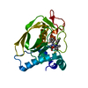
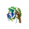
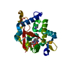
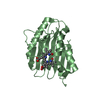
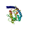
- PDB-5ej0: The vaccinia virus H3 envelope protein, a major target of neutral... -
+ Open data

- Basic information
| Entry | Database: PDB / ID: 5ej0 | ||||||
|---|---|---|---|---|---|---|---|
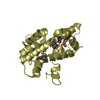
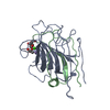
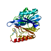
| Title | The vaccinia virus H3 envelope protein, a major target of neutralizing antibodies, exhibits a glycosyltransferase fold and binds UDP-Glucose | ||||||
 Components Components | Envelope protein H3 | ||||||
 Keywords Keywords | VIRAL PROTEIN / H3 / vaccinia virus / poxvirus / glycosyl transferase | ||||||
| Function / homology |  Function and homology information Function and homology informationmembrane => GO:0016020 / symbiont entry into host cell / viral envelope / virion attachment to host cell / virion membraneSimilarity search - Function | ||||||
| Biological species |  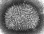 Vaccinia virus Vaccinia virus | ||||||
| Method | X-RAY DIFFRACTION / SYNCHROTRON / MAD / Resolution: 1.9 Å | ||||||
 Authors Authors | Singh, K. / Gittis, A.G. / Gitti, R.K. / Ostazesky, S.A. / Su, H.P. / Garboczi, D.N. | ||||||
 Citation Citation | Journal: J.Virol. / Year: 2016 Title: The Vaccinia Virus H3 Envelope Protein, a Major Target of Neutralizing Antibodies, Exhibits a Glycosyltransferase Fold and Binds UDP-Glucose. Authors: Singh, K. / Gittis, A.G. / Gitti, R.K. / Ostazeski, S.A. / Su, H.P. / Garboczi, D.N. | ||||||
| History |
|
- Structure visualization
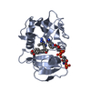
| Structure viewer | Molecule: MolmilJmol/JSmol |
|---|
- Downloads & links
-Download
| PDBx/mmCIF format | 5ej0.cif.gz | 64.2 KB | Display | PDBx/mmCIF format |
|---|---|---|---|---|
| PDB format | pdb5ej0.ent.gz | 44.3 KB | Display | PDB format |
| PDBx/mmJSON format | 5ej0.json.gz | Tree view | PDBx/mmJSON format | |
| Others |  Other downloads Other downloads |
-Validation report
| Arichive directory | https://data.pdbj.org/pub/pdb/validation_reports/ej/5ej0ftp://data.pdbj.org/pub/pdb/validation_reports/ej/5ej0 | HTTPS FTP |
|---|
-Related structure data
| Similar structure data |
|---|

-Links
 PDBj
PDBj


- Assembly
| Deposited unit | 
| ||||||||
|---|---|---|---|---|---|---|---|---|---|
| 1 |
| ||||||||
| Unit cell |
|
-Components
-Protein , 1 types, 1 molecules A
| #1: Protein | / Ag35 / Virion envelope protein p35 Mass: 27486.207 Da / Num. of mol.: 1 Source method: isolated from a genetically manipulated source Source: (gene. exp.) Vaccinia virus (strain Western Reserve)Strain: Western Reserve / Gene: VACWR101, H3L / Plasmid: pNAN / Production host:  Escherichia coli (E. coli) / References: UniProt: P07240 Escherichia coli (E. coli) / References: UniProt: P07240 |
|---|
-Non-polymers , 10 types, 116 molecules 


| #2: Chemical | ChemComp-DHL / Cysteamine Type: L-peptide linking / Mass: 77.149 Da / Num. of mol.: 1 / Source method: obtained synthetically / Formula: C2H7NS Type: L-peptide linking / Mass: 77.149 Da / Num. of mol.: 1 / Source method: obtained synthetically / Formula: C2H7NS | ||||||||||||||||
|---|---|---|---|---|---|---|---|---|---|---|---|---|---|---|---|---|---|
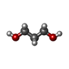
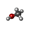
| #3: Chemical | ChemComp-PG0 / 2-(2-Methoxyethoxy)ethanol Mass: 120.147 Da / Num. of mol.: 4 / Source method: obtained synthetically / Formula: C5H12O3 / Comment: inhibitor, precipitant*YM Mass: 120.147 Da / Num. of mol.: 4 / Source method: obtained synthetically / Formula: C5H12O3 / Comment: inhibitor, precipitant*YM#4: Chemical | Glycerol Mass: 92.094 Da / Num. of mol.: 2 / Source method: obtained synthetically / Formula: C3H8O3 Mass: 92.094 Da / Num. of mol.: 2 / Source method: obtained synthetically / Formula: C3H8O3#5: Chemical | ChemComp-POL / | Propan-1-ol Mass: 60.095 Da / Num. of mol.: 1 / Source method: obtained synthetically / Formula: C3H8O Mass: 60.095 Da / Num. of mol.: 1 / Source method: obtained synthetically / Formula: C3H8O#6: Chemical | 1,3-Propanediol Mass: 76.094 Da / Num. of mol.: 2 / Source method: obtained synthetically / Formula: C3H8O2 Mass: 76.094 Da / Num. of mol.: 2 / Source method: obtained synthetically / Formula: C3H8O2#7: Chemical | ChemComp-EDO / Ethylene glycol Mass: 62.068 Da / Num. of mol.: 5 / Source method: obtained synthetically / Formula: C2H6O2 Mass: 62.068 Da / Num. of mol.: 5 / Source method: obtained synthetically / Formula: C2H6O2#8: Chemical | Ethanol Mass: 46.068 Da / Num. of mol.: 3 / Source method: obtained synthetically / Formula: C2H6O Mass: 46.068 Da / Num. of mol.: 3 / Source method: obtained synthetically / Formula: C2H6O#9: Chemical | ChemComp-MOH / Methanol Mass: 32.042 Da / Num. of mol.: 6 / Source method: obtained synthetically / Formula: CH4O Mass: 32.042 Da / Num. of mol.: 6 / Source method: obtained synthetically / Formula: CH4O#10: Chemical | ChemComp-MG / |  Mass: 24.305 Da / Num. of mol.: 1 / Source method: obtained synthetically / Formula: Mg Mass: 24.305 Da / Num. of mol.: 1 / Source method: obtained synthetically / Formula: Mg#11: Water | ChemComp-HOH / | WaterMass: 18.015 Da / Num. of mol.: 91 / Source method: isolated from a natural source / Formula: H2O |
-Details
| Sequence details | AUTHORS HAVE INDICATED THAT THE SEQUENCE IN THE UNPROT ENTRY P07240 IS INCORRECT |
|---|
-Experimental details
-Experiment
| Experiment | Method: X-RAY DIFFRACTION / Number of used crystals: 1 |
|---|
- Sample preparation
| Crystal | Density Matthews: 2.36 Å3/Da / Density % sol: 47.97 % |
|---|---|
| Crystal grow | Temperature: 298 K / Method: vapor diffusion, hanging drop / pH: 4 Details: 100 mM citric acid, 10-20% PEG 6000, 5% 1,3-propanediol and mixture of low molecular mass alcohols |
-Data collection
| Diffraction | Mean temperature: 95 K |
|---|---|
| Diffraction source | Source: SYNCHROTRON / Site: APS  / Beamline: 19-ID / Wavelength: 0.979 Å / Beamline: 19-ID / Wavelength: 0.979 Å |
| Detector | Type: ADSC QUANTUM 315 / Detector: CCD / Date: Jul 28, 2005 |
| Radiation | Monochromator: Si(111) double crystal / Protocol: SINGLE WAVELENGTH / Monochromatic (M) / Laue (L): M / Scattering type: x-ray |
| Radiation wavelength | Wavelength: 0.979 Å / Relative weight: 1 |
| Reflection | Resolution: 1.9→49.22 Å / Num. obs: 21213 / % possible obs: 99.81 % / Observed criterion σ(F): 0 / Observed criterion σ(I): -3 / Redundancy: 4.79 % / Rmerge(I) obs: 0.059 / Net I/σ(I): 18.54 |
- Processing
| Software |
| |||||||||||||||||||||||||||||||||||||||||||||||||||||||||||||||||||||||||||||||||||||||||||
|---|---|---|---|---|---|---|---|---|---|---|---|---|---|---|---|---|---|---|---|---|---|---|---|---|---|---|---|---|---|---|---|---|---|---|---|---|---|---|---|---|---|---|---|---|---|---|---|---|---|---|---|---|---|---|---|---|---|---|---|---|---|---|---|---|---|---|---|---|---|---|---|---|---|---|---|---|---|---|---|---|---|---|---|---|---|---|---|---|---|---|---|---|
| Refinement | Method to determine structure: MAD / Resolution: 1.9→49.216 Å / SU ML: 0.25 / Cross valid method: THROUGHOUT / σ(F): 1.35 / Phase error: 23.26 / Stereochemistry target values: ML
| |||||||||||||||||||||||||||||||||||||||||||||||||||||||||||||||||||||||||||||||||||||||||||
| Solvent computation | Shrinkage radii: 0.9 Å / VDW probe radii: 1.11 Å / Solvent model: FLAT BULK SOLVENT MODEL | |||||||||||||||||||||||||||||||||||||||||||||||||||||||||||||||||||||||||||||||||||||||||||
| Displacement parameters | Biso max: 97.01 Å2 / Biso mean: 33.3324 Å2 / Biso min: 11.64 Å2 | |||||||||||||||||||||||||||||||||||||||||||||||||||||||||||||||||||||||||||||||||||||||||||
| Refinement step | Cycle: final / Resolution: 1.9→49.216 Å
| |||||||||||||||||||||||||||||||||||||||||||||||||||||||||||||||||||||||||||||||||||||||||||
| Refine LS restraints |
| |||||||||||||||||||||||||||||||||||||||||||||||||||||||||||||||||||||||||||||||||||||||||||
| LS refinement shell | Refine-ID: X-RAY DIFFRACTION / Total num. of bins used: 12
|
