 Movie
Movie Controller


+ Open data

- Basic information
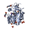
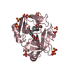
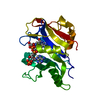
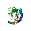
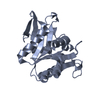
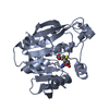
| Entry | Database: PDB / ID: 3hy6 | ||||||
|---|---|---|---|---|---|---|---|
| Title | Structure of human MTHFS with ADP | ||||||
 Components Components | 5-formyltetrahydrofolate cyclo-ligase | ||||||
 Keywords Keywords | LIGASE / antifolate / cancer / ADP / ATP-binding / Folate-binding / Magnesium / Nucleotide-binding | ||||||
| Function / homology |  Function and homology information Function and homology informationfolic acid catabolic process / 5-formyltetrahydrofolate cyclo-ligase / 5-formyltetrahydrofolate cyclo-ligase activity / folic acid-containing compound biosynthetic process / formate metabolic process / glutamate metabolic process / Metabolism of folate and pterines / tetrahydrofolate metabolic process / tetrahydrofolate interconversion / folic acid binding ...folic acid catabolic process / 5-formyltetrahydrofolate cyclo-ligase / 5-formyltetrahydrofolate cyclo-ligase activity / folic acid-containing compound biosynthetic process / formate metabolic process / glutamate metabolic process / Metabolism of folate and pterines / tetrahydrofolate metabolic process / tetrahydrofolate interconversion / folic acid binding / folic acid metabolic process / mitochondrial matrix / mitochondrion / ATP binding / metal ion binding / cytosol / cytoplasmSimilarity search - Function | ||||||
| Biological species |  Homo sapiens (human) Homo sapiens (human) | ||||||
| Method | X-RAY DIFFRACTION / SYNCHROTRON / MOLECULAR REPLACEMENT / Resolution: 2.1 Å | ||||||
 Authors Authors | Wu, D. / Li, Y. / Song, G. / Cheng, C. / Shaw, N. / Liu, Z.-J. | ||||||
 Citation Citation | Journal: Cancer Res. / Year: 2009 Title: Structural basis for the inhibition of human 5,10-methenyltetrahydrofolate synthetase by N10-substituted folate analogues Authors: Wu, D. / Li, Y. / Song, G. / Cheng, C. / Zhang, R. / Joachimiak, A. / Shaw, N. / Liu, Z.-J. | ||||||
| History |
|
- Structure visualization
| Structure viewer | Molecule: MolmilJmol/JSmol |
|---|
- Downloads & links
-Download
| PDBx/mmCIF format | 3hy6.cif.gz | 60 KB | Display | PDBx/mmCIF format |
|---|---|---|---|---|
| PDB format | pdb3hy6.ent.gz | 41.9 KB | Display | PDB format |
| PDBx/mmJSON format | 3hy6.json.gz | Tree view | PDBx/mmJSON format | |
| Others |  Other downloads Other downloads |
-Validation report
| Arichive directory | https://data.pdbj.org/pub/pdb/validation_reports/hy/3hy6ftp://data.pdbj.org/pub/pdb/validation_reports/hy/3hy6 | HTTPS FTP |
|---|
-Related structure data
| Related structure data |  3hxtSC  3hy3C  3hy4C S: Starting model for refinement C: citing same article ( |
|---|---|
| Similar structure data |


-Links
 PDBj
PDBj

- Assembly
| Deposited unit | 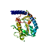
| |||||||||
|---|---|---|---|---|---|---|---|---|---|---|
| 1 |
| |||||||||
| 2 | 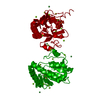
| |||||||||
| Unit cell |
| |||||||||
| Components on special symmetry positions |
|
-Components
-Protein , 1 types, 1 molecules A
| #1: Protein | / 5 / 10-methenyl-tetrahydrofolate synthetase / Methenyl-THF synthetase / MTHFS Mass: 23289.693 Da / Num. of mol.: 1 Source method: isolated from a genetically manipulated source Source: (gene. exp.) Homo sapiens (human) / Gene: MTHFS / Plasmid: pET21b / Production host:  Escherichia coli (E. coli) / Strain (production host): BL21 Escherichia coli (E. coli) / Strain (production host): BL21References: UniProt: P49914, 5-formyltetrahydrofolate cyclo-ligase |
|---|
-Non-polymers , 5 types, 164 molecules 


| #2: Chemical | Nickel Mass: 58.693 Da / Num. of mol.: 3 / Source method: obtained synthetically / Formula: Ni Mass: 58.693 Da / Num. of mol.: 3 / Source method: obtained synthetically / Formula: Ni#3: Chemical | ChemComp-MG /  Mass: 24.305 Da / Num. of mol.: 12 / Source method: obtained synthetically / Formula: Mg Mass: 24.305 Da / Num. of mol.: 12 / Source method: obtained synthetically / Formula: Mg#4: Chemical | ChemComp-PO4 / | Phosphate Mass: 94.971 Da / Num. of mol.: 1 / Source method: obtained synthetically / Formula: PO4 Mass: 94.971 Da / Num. of mol.: 1 / Source method: obtained synthetically / Formula: PO4#5: Chemical | ChemComp-ADP / | Adenosine diphosphate Mass: 427.201 Da / Num. of mol.: 1 / Source method: obtained synthetically / Formula: C10H15N5O10P2 / Comment: ADP, energy-carrying molecule*YM Mass: 427.201 Da / Num. of mol.: 1 / Source method: obtained synthetically / Formula: C10H15N5O10P2 / Comment: ADP, energy-carrying molecule*YM#6: Water | ChemComp-HOH / | WaterMass: 18.015 Da / Num. of mol.: 147 / Source method: isolated from a natural source / Formula: H2O |
|---|
-Experimental details
-Experiment
| Experiment | Method: X-RAY DIFFRACTION / Number of used crystals: 1 |
|---|
- Sample preparation
| Crystal | Density Matthews: 2.26 Å3/Da / Density % sol: 45.47 % |
|---|---|
| Crystal grow | Temperature: 298 K / Method: vapor diffusion, hanging drop / pH: 6.6 Details: 100mM HEPES, pH6.6, 20mM MgCl2.6H2O, 20mM NiCl2.6H2O, 20% (w/v) PEG 3350, VAPOR DIFFUSION, HANGING DROP, temperature 298K |
-Data collection
| Diffraction | Mean temperature: 100 K |
|---|---|
| Diffraction source | Source: SYNCHROTRON / Site: APS  / Beamline: 19-ID / Wavelength: 0.979 Å / Beamline: 19-ID / Wavelength: 0.979 Å |
| Detector | Type: ADSC QUANTUM 315r / Detector: CCD / Date: Mar 10, 2009 |
| Radiation | Protocol: SINGLE WAVELENGTH / Monochromatic (M) / Laue (L): M / Scattering type: x-ray |
| Radiation wavelength | Wavelength: 0.979 Å / Relative weight: 1 |
| Reflection | Resolution: 2.1→72.55 Å / Num. all: 12640 / Num. obs: 12552 / % possible obs: 99.3 % / Observed criterion σ(F): 2 / Observed criterion σ(I): 2 |
| Reflection shell | Resolution: 2→2.1 Å / % possible all: 99.3 |
- Processing
| Software |
| |||||||||||||||||||||||||||||||||||||||||||||||||||||||||||||||||
|---|---|---|---|---|---|---|---|---|---|---|---|---|---|---|---|---|---|---|---|---|---|---|---|---|---|---|---|---|---|---|---|---|---|---|---|---|---|---|---|---|---|---|---|---|---|---|---|---|---|---|---|---|---|---|---|---|---|---|---|---|---|---|---|---|---|---|
| Refinement | Method to determine structure: MOLECULAR REPLACEMENT Starting model: PDB ENTRY 3HXT Resolution: 2.1→46.09 Å / Cor.coef. Fo:Fc: 0.943 / Cor.coef. Fo:Fc free: 0.933 / SU B: 6.42 / SU ML: 0.169 / Cross valid method: THROUGHOUT / σ(F): 2 / ESU R: 0.278 / ESU R Free: 0.211 / Stereochemistry target values: MAXIMUM LIKELIHOOD / Details: HYDROGENS HAVE BEEN ADDED IN THE RIDING POSITIONS
| |||||||||||||||||||||||||||||||||||||||||||||||||||||||||||||||||
| Solvent computation | Ion probe radii: 0.8 Å / Shrinkage radii: 0.8 Å / VDW probe radii: 1.2 Å / Solvent model: MASK | |||||||||||||||||||||||||||||||||||||||||||||||||||||||||||||||||
| Displacement parameters | Biso mean: 35.977 Å2
| |||||||||||||||||||||||||||||||||||||||||||||||||||||||||||||||||
| Refinement step | Cycle: LAST / Resolution: 2.1→46.09 Å
| |||||||||||||||||||||||||||||||||||||||||||||||||||||||||||||||||
| Refine LS restraints |
| |||||||||||||||||||||||||||||||||||||||||||||||||||||||||||||||||
| LS refinement shell | Resolution: 2.104→2.159 Å / Total num. of bins used: 20
|
