 Movie
Movie Controller

[English] 日本語
 Yorodumi
- PDB-5e8p: The structure of the TEIPP associated altered peptide ligand Trh4... -
+ Open data

- Basic information
| Entry | Database: PDB / ID: 5e8p | ||||||
|---|---|---|---|---|---|---|---|
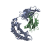
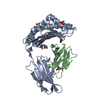
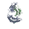
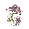
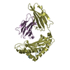
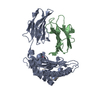
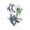
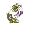
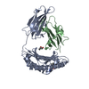
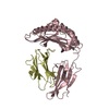
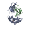
| Title | The structure of the TEIPP associated altered peptide ligand Trh4-p5NLE in complex with H-2D(b) | ||||||
 Components Components |
| ||||||
 Keywords Keywords |  IMMUNE SYSTEM / Cancer / Neo-epitope / TAP-deficiency / TEIPP / MHC-I / Sulfur-pi interactions / Non-classical peptide binding IMMUNE SYSTEM / Cancer / Neo-epitope / TAP-deficiency / TEIPP / MHC-I / Sulfur-pi interactions / Non-classical peptide binding | ||||||
| Function / homology |  Function and homology informationsphingosine N-acyltransferase / sphingoid base N-palmitoyltransferase / Sphingolipid de novo biosynthesis / sphingosine N-acyltransferase activity / sphingolipid biosynthetic process / ceramide biosynthetic process / Endosomal/Vacuolar pathway / DAP12 interactions / Antigen Presentation: Folding, assembly and peptide loading of class I MHC / ER-Phagosome pathway ...sphingosine N-acyltransferase / sphingoid base N-palmitoyltransferase / Sphingolipid de novo biosynthesis / sphingosine N-acyltransferase activity / sphingolipid biosynthetic process / ceramide biosynthetic process / Endosomal/Vacuolar pathway / DAP12 interactions / Antigen Presentation: Folding, assembly and peptide loading of class I MHC / ER-Phagosome pathway / DAP12 signaling / Immunoregulatory interactions between a Lymphoid and a non-Lymphoid cell / regulation of membrane depolarization / antigen processing and presentation of endogenous peptide antigen via MHC class I via ER pathway, TAP-dependent / cellular defense response / beta-2-microglobulin binding / Neutrophil degranulation / antigen processing and presentation of endogenous peptide antigen via MHC class I via ER pathway, TAP-independent / antigen processing and presentation of endogenous peptide antigen via MHC class Ib / lumenal side of endoplasmic reticulum membrane / peptide binding / cellular response to iron(III) ion / antigen processing and presentation of exogenous protein antigen via MHC class Ib, TAP-dependent / negative regulation of forebrain neuron differentiation / regulation of erythrocyte differentiation / peptide antigen assembly with MHC class I protein complex / response to molecule of bacterial origin / regulation of iron ion transport / MHC class I peptide loading complex / HFE-transferrin receptor complex / T cell mediated cytotoxicity / cellular response to iron ion / antigen processing and presentation of endogenous peptide antigen via MHC class I / positive regulation of T cell cytokine production / MHC class I protein complex / multicellular organismal-level iron ion homeostasis / negative regulation of neurogenesis / peptide antigen assembly with MHC class II protein complex / positive regulation of receptor-mediated endocytosis / MHC class II protein complex / cellular response to nicotine / positive regulation of T cell mediated cytotoxicity / phagocytic vesicle membrane / peptide antigen binding / positive regulation of cellular senescence / antigen processing and presentation of exogenous peptide antigen via MHC class II / negative regulation of epithelial cell proliferation / positive regulation of immune response / antimicrobial humoral immune response mediated by antimicrobial peptide / sensory perception of smell / positive regulation of T cell activation / negative regulation of neuron projection development / MHC class II protein complex binding / late endosome membrane / T cell differentiation in thymus / iron ion transport / protein refolding / antibacterial humoral response / protein homotetramerization / intracellular iron ion homeostasis / cellular response to lipopolysaccharide / defense response to Gram-negative bacterium / amyloid fibril formation / learning or memory / defense response to Gram-positive bacterium / immune response / lysosomal membrane / external side of plasma membrane / signaling receptor binding / innate immune response / protein-containing complex binding / endoplasmic reticulum membrane / structural molecule activity / Golgi apparatus / endoplasmic reticulum / protein homodimerization activity / DNA binding / extracellular space / identical protein binding / plasma membrane / cytosol Function and homology informationsphingosine N-acyltransferase / sphingoid base N-palmitoyltransferase / Sphingolipid de novo biosynthesis / sphingosine N-acyltransferase activity / sphingolipid biosynthetic process / ceramide biosynthetic process / Endosomal/Vacuolar pathway / DAP12 interactions / Antigen Presentation: Folding, assembly and peptide loading of class I MHC / ER-Phagosome pathway ...sphingosine N-acyltransferase / sphingoid base N-palmitoyltransferase / Sphingolipid de novo biosynthesis / sphingosine N-acyltransferase activity / sphingolipid biosynthetic process / ceramide biosynthetic process / Endosomal/Vacuolar pathway / DAP12 interactions / Antigen Presentation: Folding, assembly and peptide loading of class I MHC / ER-Phagosome pathway / DAP12 signaling / Immunoregulatory interactions between a Lymphoid and a non-Lymphoid cell / regulation of membrane depolarization / antigen processing and presentation of endogenous peptide antigen via MHC class I via ER pathway, TAP-dependent / cellular defense response / beta-2-microglobulin binding / Neutrophil degranulation / antigen processing and presentation of endogenous peptide antigen via MHC class I via ER pathway, TAP-independent / antigen processing and presentation of endogenous peptide antigen via MHC class Ib / lumenal side of endoplasmic reticulum membrane / peptide binding / cellular response to iron(III) ion / antigen processing and presentation of exogenous protein antigen via MHC class Ib, TAP-dependent / negative regulation of forebrain neuron differentiation / regulation of erythrocyte differentiation / peptide antigen assembly with MHC class I protein complex / response to molecule of bacterial origin / regulation of iron ion transport / MHC class I peptide loading complex / HFE-transferrin receptor complex / T cell mediated cytotoxicity / cellular response to iron ion / antigen processing and presentation of endogenous peptide antigen via MHC class I / positive regulation of T cell cytokine production / MHC class I protein complex / multicellular organismal-level iron ion homeostasis / negative regulation of neurogenesis / peptide antigen assembly with MHC class II protein complex / positive regulation of receptor-mediated endocytosis / MHC class II protein complex / cellular response to nicotine / positive regulation of T cell mediated cytotoxicity / phagocytic vesicle membrane / peptide antigen binding / positive regulation of cellular senescence / antigen processing and presentation of exogenous peptide antigen via MHC class II / negative regulation of epithelial cell proliferation / positive regulation of immune response / antimicrobial humoral immune response mediated by antimicrobial peptide / sensory perception of smell / positive regulation of T cell activation / negative regulation of neuron projection development / MHC class II protein complex binding / late endosome membrane / T cell differentiation in thymus / iron ion transport / protein refolding / antibacterial humoral response / protein homotetramerization / intracellular iron ion homeostasis / cellular response to lipopolysaccharide / defense response to Gram-negative bacterium / amyloid fibril formation / learning or memory / defense response to Gram-positive bacterium / immune response / lysosomal membrane / external side of plasma membrane / signaling receptor binding / innate immune response / protein-containing complex binding / endoplasmic reticulum membrane / structural molecule activity / Golgi apparatus / endoplasmic reticulum / protein homodimerization activity / DNA binding / extracellular space / identical protein binding / plasma membrane / cytosolSimilarity search - Function | ||||||
| Biological species |  Mus musculus (house mouse) Mus musculus (house mouse) | ||||||
| Method | X-RAY DIFFRACTION / SYNCHROTRON / MOLECULAR REPLACEMENT / Resolution: 2 Å | ||||||
 Authors Authors | Hafstrand, I. / Doorduijn, E. / Duru, A.D. / Buratto, J. / Oliveira, C.C. / Sandalova, T. / van Hall, T. / Achour, A. | ||||||
 Citation Citation | Journal: J Immunol. / Year: 2016 Title: The MHC Class I Cancer-Associated Neoepitope Trh4 Linked with Impaired Peptide Processing Induces a Unique Noncanonical TCR Conformer. Authors: Hafstrand, I. / Doorduijn, E.M. / Duru, A.D. / Buratto, J. / Oliveira, C.C. / Sandalova, T. / van Hall, T. / Achour, A. | ||||||
| History |
|
- Structure visualization
| Structure viewer | Molecule: MolmilJmol/JSmol |
|---|
- Downloads & links
-Download
| PDBx/mmCIF format | 5e8p.cif.gz | 172.2 KB | Display | PDBx/mmCIF format |
|---|---|---|---|---|
| PDB format | pdb5e8p.ent.gz | 136.7 KB | Display | PDB format |
| PDBx/mmJSON format | 5e8p.json.gz | Tree view | PDBx/mmJSON format | |
| Others |  Other downloads Other downloads |
-Validation report
| Arichive directory | https://data.pdbj.org/pub/pdb/validation_reports/e8/5e8pftp://data.pdbj.org/pub/pdb/validation_reports/e8/5e8p | HTTPS FTP |
|---|
-Related structure data
| Related structure data |  5e8nC  5e8oC 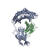 1s7uS S: Starting model for refinement C: citing same article ( |
|---|---|
| Similar structure data |


-Links
 PDBj
PDBj


- Assembly
| Deposited unit | 
| ||||||||
|---|---|---|---|---|---|---|---|---|---|
| 1 | 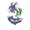
| ||||||||
| 2 | 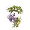
| ||||||||
| Unit cell |
|
-Components
| #1: Protein | Mass: 32087.703 Da / Num. of mol.: 2 Source method: isolated from a genetically manipulated source Source: (gene. exp.) Mus musculus (house mouse) / Gene: H2-D1 / Production host:  Escherichia coli (E. coli) / References: UniProt: P01899 Escherichia coli (E. coli) / References: UniProt: P01899#2: Protein | Beta-2 microglobulinMass: 11704.359 Da / Num. of mol.: 2 Source method: isolated from a genetically manipulated source Source: (gene. exp.) Mus musculus (house mouse) / Gene: B2m / Production host: Escherichia coli (E. coli) / References: UniProt: P01887#3: Protein/peptide | / CerS5 / LAG1 longevity assurance homolog 5 / Translocating chain-associating membrane protein ...CerS5 / LAG1 longevity assurance homolog 5 / Translocating chain-associating membrane protein homolog 4 / TRAM homolog 4Mass: 1038.371 Da / Num. of mol.: 2 / Fragment: UNP residues 379-387 / Source method: obtained synthetically / Source: (synth.) Mus musculus (house mouse) / References: UniProt: Q9D6K9, sphingosine N-acyltransferase#4: Chemical | ChemComp-SO4 / | Sulfate  Mass: 96.063 Da / Num. of mol.: 1 / Source method: obtained synthetically / Formula: SO4 Mass: 96.063 Da / Num. of mol.: 1 / Source method: obtained synthetically / Formula: SO4#5: Water | ChemComp-HOH / | Water Mass: 18.015 Da / Num. of mol.: 245 / Source method: isolated from a natural source / Formula: H2O Mass: 18.015 Da / Num. of mol.: 245 / Source method: isolated from a natural source / Formula: H2O |
|---|
-Experimental details
-Experiment
| Experiment | Method: X-RAY DIFFRACTION |
|---|
- Sample preparation
| Crystal | Density Matthews: 2.96 Å3/Da / Density % sol: 58.43 % |
|---|---|
| Crystal grow | Temperature: 277 K / Method: vapor diffusion, hanging drop / pH: 8 Details: 1.6 M ammonium sulphate, 0.1 M Tris-HCl, 0.5 M NaCl |
-Data collection
| Diffraction | Mean temperature: 100 K |
|---|---|
| Diffraction source | Source: SYNCHROTRON / Site: ESRF  / Beamline: ID14-2 / Wavelength: 0.965 Å / Beamline: ID14-2 / Wavelength: 0.965 Å |
| Detector | Type: PSI PILATUS 6M / Detector: PIXEL / Date: Mar 13, 2013 / Details: ID30A-1/MASSIF-1, PILATUS3 2M |
| Radiation | Protocol: SINGLE WAVELENGTH / Monochromatic (M) / Laue (L): M / Scattering type: x-ray |
| Radiation wavelength | Wavelength: 0.965 Å / Relative weight: 1 |
| Reflection | Resolution: 2→32.3 Å / Num. obs: 68583 / % possible obs: 96.5 % / Observed criterion σ(I): 4.2 / Redundancy: 2.5 % / Rmerge(I) obs: 0.08 / Net I/σ(I): 4.2 |
| Reflection shell | Resolution: 2→2.04 Å / Redundancy: 2.5 % / Rmerge(I) obs: 0.34 / Mean I/σ(I) obs: 1.8 / % possible all: 98.3 |
- Processing
| Software |
| |||||||||||||||||||||||||||||||||||||||||||||||||||||||||||||||||||||||||||||||||||||||||||||||||||||||||||||||||||||||||||||||||||||||||||||||||||||||||||||||||||||||||||||||
|---|---|---|---|---|---|---|---|---|---|---|---|---|---|---|---|---|---|---|---|---|---|---|---|---|---|---|---|---|---|---|---|---|---|---|---|---|---|---|---|---|---|---|---|---|---|---|---|---|---|---|---|---|---|---|---|---|---|---|---|---|---|---|---|---|---|---|---|---|---|---|---|---|---|---|---|---|---|---|---|---|---|---|---|---|---|---|---|---|---|---|---|---|---|---|---|---|---|---|---|---|---|---|---|---|---|---|---|---|---|---|---|---|---|---|---|---|---|---|---|---|---|---|---|---|---|---|---|---|---|---|---|---|---|---|---|---|---|---|---|---|---|---|---|---|---|---|---|---|---|---|---|---|---|---|---|---|---|---|---|---|---|---|---|---|---|---|---|---|---|---|---|---|---|---|---|---|
| Refinement | Method to determine structure: MOLECULAR REPLACEMENT Starting model: 1S7U Resolution: 2→31.802 Å / SU ML: 0.26 / Cross valid method: FREE R-VALUE / σ(F): 1.35 / Phase error: 27.78 / Stereochemistry target values: ML
| |||||||||||||||||||||||||||||||||||||||||||||||||||||||||||||||||||||||||||||||||||||||||||||||||||||||||||||||||||||||||||||||||||||||||||||||||||||||||||||||||||||||||||||||
| Solvent computation | Shrinkage radii: 0.9 Å / VDW probe radii: 1.11 Å / Solvent model: FLAT BULK SOLVENT MODEL | |||||||||||||||||||||||||||||||||||||||||||||||||||||||||||||||||||||||||||||||||||||||||||||||||||||||||||||||||||||||||||||||||||||||||||||||||||||||||||||||||||||||||||||||
| Refinement step | Cycle: LAST / Resolution: 2→31.802 Å
| |||||||||||||||||||||||||||||||||||||||||||||||||||||||||||||||||||||||||||||||||||||||||||||||||||||||||||||||||||||||||||||||||||||||||||||||||||||||||||||||||||||||||||||||
| Refine LS restraints |
| |||||||||||||||||||||||||||||||||||||||||||||||||||||||||||||||||||||||||||||||||||||||||||||||||||||||||||||||||||||||||||||||||||||||||||||||||||||||||||||||||||||||||||||||
| LS refinement shell |
|
