 Movie
Movie Controller


+ Open data

- Basic information
| Entry | Database: PDB / ID: 3cc5 | ||||||
|---|---|---|---|---|---|---|---|
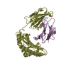
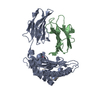
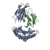
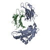
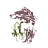
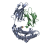
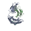
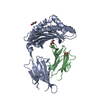
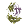
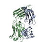
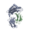
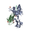
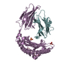
| Title | H-2Db complex with human gp100 | ||||||
 Components Components |
| ||||||
 Keywords Keywords |  IMMUNE SYSTEM / murine MHC / Glycoprotein / Immune response / Membrane / MHC I / Transmembrane / Immunoglobulin domain / Secreted / Melanin biosynthesis IMMUNE SYSTEM / murine MHC / Glycoprotein / Immune response / Membrane / MHC I / Transmembrane / Immunoglobulin domain / Secreted / Melanin biosynthesis | ||||||
| Function / homology |  Function and homology information Function and homology informationpositive regulation of melanin biosynthetic process / cis-Golgi network membrane / melanin biosynthetic process / melanosome membrane / multivesicular body, internal vesicle / melanosome organization / multivesicular body membrane / Endosomal/Vacuolar pathway / DAP12 interactions / Antigen Presentation: Folding, assembly and peptide loading of class I MHC ...positive regulation of melanin biosynthetic process / cis-Golgi network membrane / melanin biosynthetic process / melanosome membrane / multivesicular body, internal vesicle / melanosome organization / multivesicular body membrane / Endosomal/Vacuolar pathway / DAP12 interactions / Antigen Presentation: Folding, assembly and peptide loading of class I MHC / ER-Phagosome pathway / DAP12 signaling / Immunoregulatory interactions between a Lymphoid and a non-Lymphoid cell / regulation of membrane depolarization / antigen processing and presentation of endogenous peptide antigen via MHC class I via ER pathway, TAP-dependent / cellular defense response / beta-2-microglobulin binding / Neutrophil degranulation / antigen processing and presentation of endogenous peptide antigen via MHC class I via ER pathway, TAP-independent / antigen processing and presentation of endogenous peptide antigen via MHC class Ib / lumenal side of endoplasmic reticulum membrane / peptide binding / cellular response to iron(III) ion / antigen processing and presentation of exogenous protein antigen via MHC class Ib, TAP-dependent / negative regulation of forebrain neuron differentiation / regulation of erythrocyte differentiation / peptide antigen assembly with MHC class I protein complex / response to molecule of bacterial origin / regulation of iron ion transport / MHC class I peptide loading complex / HFE-transferrin receptor complex / T cell mediated cytotoxicity / cellular response to iron ion / antigen processing and presentation of endogenous peptide antigen via MHC class I / positive regulation of T cell cytokine production / MHC class I protein complex / multicellular organismal-level iron ion homeostasis / negative regulation of neurogenesis / peptide antigen assembly with MHC class II protein complex / positive regulation of receptor-mediated endocytosis / positive regulation of T cell mediated cytotoxicity / MHC class II protein complex / cellular response to nicotine / phagocytic vesicle membrane / peptide antigen binding / positive regulation of cellular senescence / antigen processing and presentation of exogenous peptide antigen via MHC class II / negative regulation of epithelial cell proliferation / positive regulation of immune response / antimicrobial humoral immune response mediated by antimicrobial peptide / melanosome / sensory perception of smell / positive regulation of T cell activation / negative regulation of neuron projection development / MHC class II protein complex binding / late endosome membrane / T cell differentiation in thymus / iron ion transport / protein refolding / antibacterial humoral response / protein homotetramerization / intracellular iron ion homeostasis / cellular response to lipopolysaccharide / defense response to Gram-negative bacterium / amyloid fibril formation / learning or memory / defense response to Gram-positive bacterium / immune response / lysosomal membrane / external side of plasma membrane / signaling receptor binding / innate immune response / protein-containing complex binding / endoplasmic reticulum membrane / structural molecule activity / Golgi apparatus / endoplasmic reticulum / protein homodimerization activity / extracellular space / extracellular exosome / identical protein binding / plasma membrane / cytosolSimilarity search - Function | ||||||
| Biological species |  Mus musculus (house mouse) Mus musculus (house mouse) | ||||||
| Method | X-RAY DIFFRACTION / SYNCHROTRON / MOLECULAR REPLACEMENT / Resolution: 1.91 Å | ||||||
 Authors Authors | Badia-Martinez, D. / Achour, A. | ||||||
 Citation Citation | Journal: Cancer Res. / Year: 2009 Title: Design of agonistic altered peptides for the robust induction of CTL directed towards H-2Db in complex with the melanoma-associated epitope gp100. Authors: van Stipdonk, M.J. / Badia-Martinez, D. / Sluijter, M. / Offringa, R. / van Hall, T. / Achour, A. | ||||||
| History |
|
- Structure visualization
| Structure viewer | Molecule: MolmilJmol/JSmol |
|---|
- Downloads & links
-Download
| PDBx/mmCIF format | 3cc5.cif.gz | 182.5 KB | Display | PDBx/mmCIF format |
|---|---|---|---|---|
| PDB format | pdb3cc5.ent.gz | 145.1 KB | Display | PDB format |
| PDBx/mmJSON format | 3cc5.json.gz | Tree view | PDBx/mmJSON format | |
| Others |  Other downloads Other downloads |
-Validation report
| Arichive directory | https://data.pdbj.org/pub/pdb/validation_reports/cc/3cc5ftp://data.pdbj.org/pub/pdb/validation_reports/cc/3cc5 | HTTPS FTP |
|---|
-Related structure data
| Related structure data | 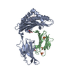 3cchC  3ch1C 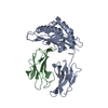 1n5aS S: Starting model for refinement C: citing same article ( |
|---|---|
| Similar structure data |


-Links
 PDBj
PDBj


- Assembly
| Deposited unit | 
| |||||||||||||||||||||||||||||||||||||||||||||||||||||||||||||||||||||||||||||||||||||||||||||||||||||||||||||||||||||||||||||||||||||||||||||||||||||||||||||||||||||||||||||||||||||||||||||||||||||||||||||||||||||||||||||||
|---|---|---|---|---|---|---|---|---|---|---|---|---|---|---|---|---|---|---|---|---|---|---|---|---|---|---|---|---|---|---|---|---|---|---|---|---|---|---|---|---|---|---|---|---|---|---|---|---|---|---|---|---|---|---|---|---|---|---|---|---|---|---|---|---|---|---|---|---|---|---|---|---|---|---|---|---|---|---|---|---|---|---|---|---|---|---|---|---|---|---|---|---|---|---|---|---|---|---|---|---|---|---|---|---|---|---|---|---|---|---|---|---|---|---|---|---|---|---|---|---|---|---|---|---|---|---|---|---|---|---|---|---|---|---|---|---|---|---|---|---|---|---|---|---|---|---|---|---|---|---|---|---|---|---|---|---|---|---|---|---|---|---|---|---|---|---|---|---|---|---|---|---|---|---|---|---|---|---|---|---|---|---|---|---|---|---|---|---|---|---|---|---|---|---|---|---|---|---|---|---|---|---|---|---|---|---|---|---|---|---|---|---|---|---|---|---|---|---|---|---|---|---|---|---|
| 1 | 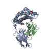
| |||||||||||||||||||||||||||||||||||||||||||||||||||||||||||||||||||||||||||||||||||||||||||||||||||||||||||||||||||||||||||||||||||||||||||||||||||||||||||||||||||||||||||||||||||||||||||||||||||||||||||||||||||||||||||||||
| 2 | 
| |||||||||||||||||||||||||||||||||||||||||||||||||||||||||||||||||||||||||||||||||||||||||||||||||||||||||||||||||||||||||||||||||||||||||||||||||||||||||||||||||||||||||||||||||||||||||||||||||||||||||||||||||||||||||||||||
| Unit cell |
| |||||||||||||||||||||||||||||||||||||||||||||||||||||||||||||||||||||||||||||||||||||||||||||||||||||||||||||||||||||||||||||||||||||||||||||||||||||||||||||||||||||||||||||||||||||||||||||||||||||||||||||||||||||||||||||||
| Noncrystallographic symmetry (NCS) | NCS domain:
NCS domain segments:
|