 Movie
Movie Controller

[English] 日本語
 Yorodumi
- PDB-5duu: Crystal structure of the human galectin-4 N-terminal carbohydrate... -
+ Open data

- Basic information
| Entry | Database: PDB / ID: 5duu | ||||||
|---|---|---|---|---|---|---|---|
| Title | Crystal structure of the human galectin-4 N-terminal carbohydrate recognition domain in complex with glycerol | ||||||
 Components Components | Galectin-4 | ||||||
 Keywords Keywords | SUGAR BINDING PROTEIN / galectin-4 / lectin / glycerol / sugar-binding protein | ||||||
| Function / homology |  Function and homology information Function and homology informationantibacterial peptide biosynthetic process / galactoside binding / carbohydrate binding / collagen-containing extracellular matrix / cell adhesion / extracellular space / plasma membrane / cytosolSimilarity search - Function | ||||||
| Biological species |  Homo sapiens (human) Homo sapiens (human) | ||||||
| Method | X-RAY DIFFRACTION / SYNCHROTRON / MOLECULAR REPLACEMENT / Resolution: 2 Å | ||||||
 Authors Authors | Bum-Erdene, K. / Blanchard, H. | ||||||
| Funding support |  Australia, 1items Australia, 1items
| ||||||
 Citation Citation | Journal: Sci Rep / Year: 2016 Title: Structural characterisation of human galectin-4 N-terminal carbohydrate recognition domain in complex with glycerol, lactose, 3'-sulfo-lactose, and 2'-fucosyllactose. Authors: Bum-Erdene, K. / Leffler, H. / Nilsson, U.J. / Blanchard, H. | ||||||
| History |
|
- Structure visualization
| Structure viewer | Molecule: MolmilJmol/JSmol |
|---|
- Downloads & links
-Download
| PDBx/mmCIF format | 5duu.cif.gz | 129.4 KB | Display | PDBx/mmCIF format |
|---|---|---|---|---|
| PDB format | pdb5duu.ent.gz | 100.1 KB | Display | PDB format |
| PDBx/mmJSON format | 5duu.json.gz | Tree view | PDBx/mmJSON format | |
| Others |  Other downloads Other downloads |
-Validation report
| Arichive directory | https://data.pdbj.org/pub/pdb/validation_reports/du/5duuftp://data.pdbj.org/pub/pdb/validation_reports/du/5duu | HTTPS FTP |
|---|
-Related structure data
| Related structure data | 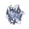 5duvC 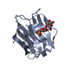 5duwC  5duxC  3i8tS C: citing same article ( S: Starting model for refinement |
|---|---|
| Similar structure data |


-Links
 PDBj
PDBj- Assembly
| Deposited unit | 
| ||||||||||||||||||||||||||||||||||||||||||||||||||||||||||||||||||||||||||||||||||||||||||||||||||||||||||||||||||||||||||||||||||||||||||||||||||||||
|---|---|---|---|---|---|---|---|---|---|---|---|---|---|---|---|---|---|---|---|---|---|---|---|---|---|---|---|---|---|---|---|---|---|---|---|---|---|---|---|---|---|---|---|---|---|---|---|---|---|---|---|---|---|---|---|---|---|---|---|---|---|---|---|---|---|---|---|---|---|---|---|---|---|---|---|---|---|---|---|---|---|---|---|---|---|---|---|---|---|---|---|---|---|---|---|---|---|---|---|---|---|---|---|---|---|---|---|---|---|---|---|---|---|---|---|---|---|---|---|---|---|---|---|---|---|---|---|---|---|---|---|---|---|---|---|---|---|---|---|---|---|---|---|---|---|---|---|---|---|---|---|
| 1 | 
| ||||||||||||||||||||||||||||||||||||||||||||||||||||||||||||||||||||||||||||||||||||||||||||||||||||||||||||||||||||||||||||||||||||||||||||||||||||||
| 2 | 
| ||||||||||||||||||||||||||||||||||||||||||||||||||||||||||||||||||||||||||||||||||||||||||||||||||||||||||||||||||||||||||||||||||||||||||||||||||||||
| 3 | 
| ||||||||||||||||||||||||||||||||||||||||||||||||||||||||||||||||||||||||||||||||||||||||||||||||||||||||||||||||||||||||||||||||||||||||||||||||||||||
| 4 | 
| ||||||||||||||||||||||||||||||||||||||||||||||||||||||||||||||||||||||||||||||||||||||||||||||||||||||||||||||||||||||||||||||||||||||||||||||||||||||
| Unit cell |
| ||||||||||||||||||||||||||||||||||||||||||||||||||||||||||||||||||||||||||||||||||||||||||||||||||||||||||||||||||||||||||||||||||||||||||||||||||||||
| Noncrystallographic symmetry (NCS) | NCS domain:
NCS domain segments: Component-ID: 0 / Beg auth comp-ID: PRO / Beg label comp-ID: PRO / Refine code: 0
NCS ensembles :
|
-Components
| #1: Protein | / Gal-4 / Antigen NY-CO-27 / L-36 lactose-binding protein / L36LBP / Lactose-binding lectin 4 Mass: 17473.912 Da / Num. of mol.: 4 Fragment: N-terminal carbohydrate recognition domain (UNP residues 1-155) Source method: isolated from a genetically manipulated source Source: (gene. exp.) Homo sapiens (human) / Gene: LGALS4 / Production host:  Escherichia coli (E. coli) / References: UniProt: P56470 Escherichia coli (E. coli) / References: UniProt: P56470#2: Chemical | ChemComp-GOL / Glycerol  Mass: 92.094 Da / Num. of mol.: 4 / Source method: obtained synthetically / Formula: C3H8O3 Mass: 92.094 Da / Num. of mol.: 4 / Source method: obtained synthetically / Formula: C3H8O3#3: Water | ChemComp-HOH / | Water Mass: 18.015 Da / Num. of mol.: 306 / Source method: isolated from a natural source / Formula: H2O Mass: 18.015 Da / Num. of mol.: 306 / Source method: isolated from a natural source / Formula: H2O |
|---|
-Experimental details
-Experiment
| Experiment | Method: X-RAY DIFFRACTION / Number of used crystals: 1 |
|---|
- Sample preparation
| Crystal | Density Matthews: 1.89 Å3/Da / Density % sol: 34.86 % |
|---|---|
| Crystal grow | Temperature: 293.15 K / Method: vapor diffusion, hanging drop / pH: 8 / Details: 3.5 M Sodium formate, 0.1 M Tris |
-Data collection
| Diffraction | Mean temperature: 100 K | |||||||||||||||||||||||||||
|---|---|---|---|---|---|---|---|---|---|---|---|---|---|---|---|---|---|---|---|---|---|---|---|---|---|---|---|---|
| Diffraction source | Source: SYNCHROTRON / Site: Australian Synchrotron / Beamline: MX2 / Wavelength: 0.9537 Å | |||||||||||||||||||||||||||
| Detector | Type: ADSC QUANTUM 315r / Detector: CCD / Date: Mar 19, 2014 | |||||||||||||||||||||||||||
| Radiation | Protocol: SINGLE WAVELENGTH / Monochromatic (M) / Laue (L): M / Scattering type: x-ray | |||||||||||||||||||||||||||
| Radiation wavelength | Wavelength: 0.9537 Å / Relative weight: 1 | |||||||||||||||||||||||||||
| Reflection | Resolution: 2→45.68 Å / Num. obs: 35098 / % possible obs: 99.5 % / Redundancy: 4.5 % / CC1/2: 0.998 / Rmerge(I) obs: 0.074 / Rpim(I) all: 0.039 / Net I/σ(I): 15.8 / Num. measured all: 159500 / Scaling rejects: 3 | |||||||||||||||||||||||||||
| Reflection shell | Diffraction-ID: 1 / Rejects: 0
|
- Processing
| Software |
| |||||||||||||||||||||||||||||||||||||||||||||||||||||||||||||||||||||||||||
|---|---|---|---|---|---|---|---|---|---|---|---|---|---|---|---|---|---|---|---|---|---|---|---|---|---|---|---|---|---|---|---|---|---|---|---|---|---|---|---|---|---|---|---|---|---|---|---|---|---|---|---|---|---|---|---|---|---|---|---|---|---|---|---|---|---|---|---|---|---|---|---|---|---|---|---|---|
| Refinement | Method to determine structure: MOLECULAR REPLACEMENT Starting model: 3I8T Resolution: 2→45.68 Å / Cor.coef. Fo:Fc: 0.947 / Cor.coef. Fo:Fc free: 0.915 / WRfactor Rfree: 0.2014 / WRfactor Rwork: 0.1682 / FOM work R set: 0.8463 / SU B: 4.273 / SU ML: 0.122 / SU R Cruickshank DPI: 0.2174 / SU Rfree: 0.1714 / Cross valid method: THROUGHOUT / σ(F): 0 / ESU R: 0.217 / ESU R Free: 0.171 / Stereochemistry target values: MAXIMUM LIKELIHOOD Details: The authors state that the electron density currently modelled as water molecules A301, B301 and D301, which are coordinated by residues ASP72, GLY70 and PHE68 shows potential for occupation ...Details: The authors state that the electron density currently modelled as water molecules A301, B301 and D301, which are coordinated by residues ASP72, GLY70 and PHE68 shows potential for occupation by NA+ ions, which is present in the crystallisation conditions.
| |||||||||||||||||||||||||||||||||||||||||||||||||||||||||||||||||||||||||||
| Solvent computation | Ion probe radii: 0.8 Å / Shrinkage radii: 0.8 Å / VDW probe radii: 1.2 Å / Solvent model: MASK | |||||||||||||||||||||||||||||||||||||||||||||||||||||||||||||||||||||||||||
| Displacement parameters | Biso max: 60.42 Å2 / Biso mean: 18.537 Å2 / Biso min: 6.25 Å2
| |||||||||||||||||||||||||||||||||||||||||||||||||||||||||||||||||||||||||||
| Refinement step | Cycle: final / Resolution: 2→45.68 Å
| |||||||||||||||||||||||||||||||||||||||||||||||||||||||||||||||||||||||||||
| Refine LS restraints |
| |||||||||||||||||||||||||||||||||||||||||||||||||||||||||||||||||||||||||||
| Refine LS restraints NCS | Refine-ID: X-RAY DIFFRACTION / Type: interatomic distance / Weight position: 0.05
| |||||||||||||||||||||||||||||||||||||||||||||||||||||||||||||||||||||||||||
| LS refinement shell | Resolution: 2.002→2.054 Å / Total num. of bins used: 20
|
