 Movie
Movie Controller


+ Open data

- Basic information
| Entry | Database: PDB / ID: 5jp5 | ||||||
|---|---|---|---|---|---|---|---|
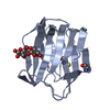
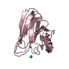
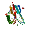
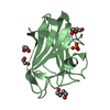
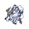
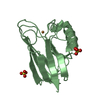
| Title | Crystal structure of rat Galectin 5 | ||||||
 Components Components | Galectin-5 | ||||||
 Keywords Keywords | SUGAR BINDING PROTEIN / lectin carbohydrate recognition jellyroll topology | ||||||
| Function / homology |  Function and homology informationlactose binding / galactoside binding / negative regulation of CD4-positive, alpha-beta T cell proliferation / positive regulation of T cell apoptotic process / negative regulation of type II interferon production / carbohydrate binding / positive regulation of gene expression / nucleus / cytosol Function and homology informationlactose binding / galactoside binding / negative regulation of CD4-positive, alpha-beta T cell proliferation / positive regulation of T cell apoptotic process / negative regulation of type II interferon production / carbohydrate binding / positive regulation of gene expression / nucleus / cytosolSimilarity search - Function | ||||||
| Biological species |  Rattus norvegicus (Norway rat) Rattus norvegicus (Norway rat) | ||||||
| Method | X-RAY DIFFRACTION / SYNCHROTRON / MOLECULAR REPLACEMENT / Resolution: 1.7 Å | ||||||
 Authors Authors | Ruiz, F.M. / Romero, A. | ||||||
| Funding support |  Spain, 1items Spain, 1items
| ||||||
 Citation Citation | Journal: Biomolecules / Year: 2021 Title: Structural Characterization of Rat Galectin-5, an N-Tailed Monomeric Proto-Type-like Galectin. Authors: Ruiz, F.M. / Medrano, F.J. / Ludwig, A.K. / Kaltner, H. / Shilova, N.V. / Bovin, N.V. / Gabius, H.J. / Romero, A. | ||||||
| History |
|
- Structure visualization
| Structure viewer | Molecule: MolmilJmol/JSmol |
|---|
- Downloads & links
-Download
| PDBx/mmCIF format | 5jp5.cif.gz | 194.8 KB | Display | PDBx/mmCIF format |
|---|---|---|---|---|
| PDB format | pdb5jp5.ent.gz | 155.7 KB | Display | PDB format |
| PDBx/mmJSON format | 5jp5.json.gz | Tree view | PDBx/mmJSON format | |
| Others |  Other downloads Other downloads |
-Validation report
| Arichive directory | https://data.pdbj.org/pub/pdb/validation_reports/jp/5jp5ftp://data.pdbj.org/pub/pdb/validation_reports/jp/5jp5 | HTTPS FTP |
|---|
-Related structure data
| Related structure data | 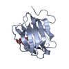 5jpgC  7p8hC 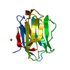 3nv1S S: Starting model for refinement C: citing same article ( |
|---|---|
| Similar structure data |


-Links
 PDBj
PDBj- Assembly
| Deposited unit | 
| ||||||||
|---|---|---|---|---|---|---|---|---|---|
| 1 | 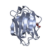
| ||||||||
| 2 | 
| ||||||||
| 3 | 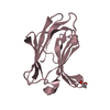
| ||||||||
| 4 | 
| ||||||||
| 5 | 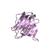
| ||||||||
| 6 | 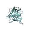
| ||||||||
| Unit cell |
|
-Components
| #1: Protein | / Gal-5 / RL-18 Mass: 16212.335 Da / Num. of mol.: 6 Source method: isolated from a genetically manipulated source Source: (gene. exp.) Rattus norvegicus (Norway rat) / Gene: Lgals5 / Production host:  Escherichia coli (E. coli) / References: UniProt: P47967 Escherichia coli (E. coli) / References: UniProt: P47967#2: Chemical | ChemComp-PEG / Diethylene glycol  Mass: 106.120 Da / Num. of mol.: 4 / Source method: obtained synthetically / Formula: C4H10O3 Mass: 106.120 Da / Num. of mol.: 4 / Source method: obtained synthetically / Formula: C4H10O3#3: Water | ChemComp-HOH / | Water Mass: 18.015 Da / Num. of mol.: 849 / Source method: isolated from a natural source / Formula: H2O Mass: 18.015 Da / Num. of mol.: 849 / Source method: isolated from a natural source / Formula: H2O |
|---|
-Experimental details
-Experiment
| Experiment | Method: X-RAY DIFFRACTION / Number of used crystals: 1 |
|---|
- Sample preparation
| Crystal | Density Matthews: 2.23 Å3/Da / Density % sol: 44.95 % |
|---|---|
| Crystal grow | Temperature: 295 K / Method: vapor diffusion, sitting drop / Details: 10% PEG 8000, 100 mM Tris pH 7.0, 200 mM MgCl2 |
-Data collection
| Diffraction | Mean temperature: 100 K |
|---|---|
| Diffraction source | Source: SYNCHROTRON / Site: ESRF  / Beamline: BM14 / Wavelength: 0.95372 Å / Beamline: BM14 / Wavelength: 0.95372 Å |
| Detector | Type: MAR CCD 130 mm / Detector: CCD / Date: May 27, 2007 |
| Radiation | Protocol: SINGLE WAVELENGTH / Monochromatic (M) / Laue (L): M / Scattering type: x-ray |
| Radiation wavelength | Wavelength: 0.95372 Å / Relative weight: 1 |
| Reflection | Resolution: 1.7→33.03 Å / Num. obs: 92950 / % possible obs: 99.75 % / Redundancy: 4.1 % / CC1/2: 0.99 / Rmerge(I) obs: 0.057 / Net I/σ(I): 14.01 |
| Reflection shell | Resolution: 1.7→1.76 Å / Redundancy: 3.7 % / Rmerge(I) obs: 0.45 / Mean I/σ(I) obs: 2.8 / % possible all: 99.21 |
- Processing
| Software |
| |||||||||||||||||||||||||||||||||||||||||||||||||||||||||||||||||||||||||||||||||||||||||||||||||||||||||||||||||||||||||||||||||||||||||||||||||||||||||||||||||||||||||||||||||||||||||||||||||||||||||||||||||||||||||
|---|---|---|---|---|---|---|---|---|---|---|---|---|---|---|---|---|---|---|---|---|---|---|---|---|---|---|---|---|---|---|---|---|---|---|---|---|---|---|---|---|---|---|---|---|---|---|---|---|---|---|---|---|---|---|---|---|---|---|---|---|---|---|---|---|---|---|---|---|---|---|---|---|---|---|---|---|---|---|---|---|---|---|---|---|---|---|---|---|---|---|---|---|---|---|---|---|---|---|---|---|---|---|---|---|---|---|---|---|---|---|---|---|---|---|---|---|---|---|---|---|---|---|---|---|---|---|---|---|---|---|---|---|---|---|---|---|---|---|---|---|---|---|---|---|---|---|---|---|---|---|---|---|---|---|---|---|---|---|---|---|---|---|---|---|---|---|---|---|---|---|---|---|---|---|---|---|---|---|---|---|---|---|---|---|---|---|---|---|---|---|---|---|---|---|---|---|---|---|---|---|---|---|---|---|---|---|---|---|---|---|---|---|---|---|---|---|---|---|
| Refinement | Method to determine structure: MOLECULAR REPLACEMENT Starting model: 3NV1 Resolution: 1.7→33.027 Å / SU ML: 0.17 / Cross valid method: FREE R-VALUE / σ(F): 1.36 / Phase error: 25.14
| |||||||||||||||||||||||||||||||||||||||||||||||||||||||||||||||||||||||||||||||||||||||||||||||||||||||||||||||||||||||||||||||||||||||||||||||||||||||||||||||||||||||||||||||||||||||||||||||||||||||||||||||||||||||||
| Solvent computation | Shrinkage radii: 0.9 Å / VDW probe radii: 1.11 Å | |||||||||||||||||||||||||||||||||||||||||||||||||||||||||||||||||||||||||||||||||||||||||||||||||||||||||||||||||||||||||||||||||||||||||||||||||||||||||||||||||||||||||||||||||||||||||||||||||||||||||||||||||||||||||
| Refinement step | Cycle: LAST / Resolution: 1.7→33.027 Å
| |||||||||||||||||||||||||||||||||||||||||||||||||||||||||||||||||||||||||||||||||||||||||||||||||||||||||||||||||||||||||||||||||||||||||||||||||||||||||||||||||||||||||||||||||||||||||||||||||||||||||||||||||||||||||
| Refine LS restraints |
| |||||||||||||||||||||||||||||||||||||||||||||||||||||||||||||||||||||||||||||||||||||||||||||||||||||||||||||||||||||||||||||||||||||||||||||||||||||||||||||||||||||||||||||||||||||||||||||||||||||||||||||||||||||||||
| LS refinement shell |
|
