 Movie
Movie Controller

[English] 日本語
 Yorodumi
- PDB-5duv: Crystal structure of the human galectin-4 N-terminal carbohydrate... -
+ Open data

- Basic information
| Entry | Database: PDB / ID: 5duv | |||||||||
|---|---|---|---|---|---|---|---|---|---|---|
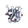
| Title | Crystal structure of the human galectin-4 N-terminal carbohydrate recognition domain in complex with lactose | |||||||||
 Components Components | Galectin-4 | |||||||||
 Keywords Keywords | SUGAR BINDING PROTEIN / galectin-4 / lectin / lactose / sugar-binding protein | |||||||||
| Function / homology |  Function and homology information Function and homology informationantibacterial peptide biosynthetic process / galactoside binding / carbohydrate binding / collagen-containing extracellular matrix / cell adhesion / extracellular space / plasma membrane / cytosolSimilarity search - Function | |||||||||
| Biological species |  Homo sapiens (human) Homo sapiens (human) | |||||||||
| Method | X-RAY DIFFRACTION / SYNCHROTRON / MOLECULAR REPLACEMENT / Resolution: 1.9 Å | |||||||||
 Authors Authors | Bum-Erdene, K. / Blanchard, H. | |||||||||
| Funding support |  Australia, 1items Australia, 1items
| |||||||||
 Citation Citation | Journal: Sci Rep / Year: 2016 Title: Structural characterisation of human galectin-4 N-terminal carbohydrate recognition domain in complex with glycerol, lactose, 3'-sulfo-lactose, and 2'-fucosyllactose. Authors: Bum-Erdene, K. / Leffler, H. / Nilsson, U.J. / Blanchard, H. | |||||||||
| History |
|
- Structure visualization
| Structure viewer | Molecule: MolmilJmol/JSmol |
|---|
- Downloads & links
-Download
| PDBx/mmCIF format | 5duv.cif.gz | 135.9 KB | Display | PDBx/mmCIF format |
|---|---|---|---|---|
| PDB format | pdb5duv.ent.gz | 104 KB | Display | PDB format |
| PDBx/mmJSON format | 5duv.json.gz | Tree view | PDBx/mmJSON format | |
| Others |  Other downloads Other downloads |
-Validation report
| Arichive directory | https://data.pdbj.org/pub/pdb/validation_reports/du/5duvftp://data.pdbj.org/pub/pdb/validation_reports/du/5duv | HTTPS FTP |
|---|
-Related structure data
| Related structure data | 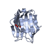 5duuC 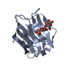 5duwC  5duxC  3i8tS C: citing same article ( S: Starting model for refinement |
|---|---|
| Similar structure data |


-Links
 PDBj
PDBj


- Assembly
| Deposited unit | 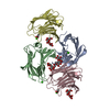
| ||||||||
|---|---|---|---|---|---|---|---|---|---|
| 1 | 
| ||||||||
| 2 | 
| ||||||||
| 3 | 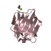
| ||||||||
| 4 | 
| ||||||||
| Unit cell |
|
-Components
| #1: Protein | / Gal-4 / Antigen NY-CO-27 / L-36 lactose-binding protein / L36LBP / Lactose-binding lectin 4 Mass: 17473.912 Da / Num. of mol.: 4 Fragment: N-terminal carbohydrate recognition domain (UNP residues 1-155) Source method: isolated from a genetically manipulated source Source: (gene. exp.) Homo sapiens (human) / Gene: LGALS4 / Production host:  Escherichia coli (E. coli) / References: UniProt: P56470 Escherichia coli (E. coli) / References: UniProt: P56470#2: Polysaccharide | beta-D-galactopyranose-(1-4)-beta-D-glucopyranose / beta-lactose   , Oligosaccharide / Class: Nutrient / Mass: 342.297 Da / Num. of mol.: 4 , Oligosaccharide / Class: Nutrient / Mass: 342.297 Da / Num. of mol.: 4Source method: isolated from a genetically manipulated source Details: oligosaccharide / References: beta-lactose #3: Chemical | ChemComp-CA /   Mass: 40.078 Da / Num. of mol.: 4 / Source method: obtained synthetically / Formula: Ca Mass: 40.078 Da / Num. of mol.: 4 / Source method: obtained synthetically / Formula: Ca#4: Chemical | Acetate  Mass: 59.044 Da / Num. of mol.: 2 / Source method: obtained synthetically / Formula: C2H3O2 Mass: 59.044 Da / Num. of mol.: 2 / Source method: obtained synthetically / Formula: C2H3O2#5: Water | ChemComp-HOH / | Water Mass: 18.015 Da / Num. of mol.: 380 / Source method: isolated from a natural source / Formula: H2O Mass: 18.015 Da / Num. of mol.: 380 / Source method: isolated from a natural source / Formula: H2O |
|---|
-Experimental details
-Experiment
| Experiment | Method: X-RAY DIFFRACTION / Number of used crystals: 1 |
|---|
- Sample preparation
| Crystal | Density Matthews: 1.88 Å3/Da / Density % sol: 34.52 % |
|---|---|
| Crystal grow | Temperature: 293.15 K / Method: vapor diffusion, hanging drop / pH: 7.2 / Details: 0.2 M calcium acetate, 20 % w/v PEG 3350 |
-Data collection
| Diffraction | Mean temperature: 100 K | |||||||||||||||||||||||||||
|---|---|---|---|---|---|---|---|---|---|---|---|---|---|---|---|---|---|---|---|---|---|---|---|---|---|---|---|---|
| Diffraction source | Source: SYNCHROTRON / Site: Australian Synchrotron / Beamline: MX1 / Wavelength: 0.9537 Å | |||||||||||||||||||||||||||
| Detector | Type: ADSC QUANTUM 210r / Detector: CCD / Date: Oct 15, 2013 | |||||||||||||||||||||||||||
| Radiation | Protocol: SINGLE WAVELENGTH / Monochromatic (M) / Laue (L): M / Scattering type: x-ray | |||||||||||||||||||||||||||
| Radiation wavelength | Wavelength: 0.9537 Å / Relative weight: 1 | |||||||||||||||||||||||||||
| Reflection | Resolution: 1.9→37.24 Å / Num. obs: 40168 / % possible obs: 98.1 % / Redundancy: 4 % / CC1/2: 0.997 / Rmerge(I) obs: 0.078 / Rpim(I) all: 0.044 / Net I/σ(I): 12.2 / Num. measured all: 161697 / Scaling rejects: 168 | |||||||||||||||||||||||||||
| Reflection shell | Diffraction-ID: 1 / Rejects: 0
|
- Processing
| Software |
| |||||||||||||||||||||||||||||||||||||||||||||||||||||||||||||||||||||||||||
|---|---|---|---|---|---|---|---|---|---|---|---|---|---|---|---|---|---|---|---|---|---|---|---|---|---|---|---|---|---|---|---|---|---|---|---|---|---|---|---|---|---|---|---|---|---|---|---|---|---|---|---|---|---|---|---|---|---|---|---|---|---|---|---|---|---|---|---|---|---|---|---|---|---|---|---|---|
| Refinement | Method to determine structure: MOLECULAR REPLACEMENT Starting model: 3I8T Resolution: 1.9→37.24 Å / Cor.coef. Fo:Fc: 0.963 / Cor.coef. Fo:Fc free: 0.947 / WRfactor Rfree: 0.1926 / WRfactor Rwork: 0.1506 / FOM work R set: 0.8706 / SU B: 3.264 / SU ML: 0.095 / SU R Cruickshank DPI: 0.1577 / SU Rfree: 0.1396 / Cross valid method: THROUGHOUT / σ(F): 0 / ESU R: 0.158 / ESU R Free: 0.14 / Stereochemistry target values: MAXIMUM LIKELIHOOD Details: HYDROGENS HAVE BEEN ADDED IN THE RIDING POSITIONS U VALUES : REFINED INDIVIDUALLY
| |||||||||||||||||||||||||||||||||||||||||||||||||||||||||||||||||||||||||||
| Solvent computation | Ion probe radii: 0.8 Å / Shrinkage radii: 0.8 Å / VDW probe radii: 1.2 Å / Solvent model: MASK | |||||||||||||||||||||||||||||||||||||||||||||||||||||||||||||||||||||||||||
| Displacement parameters | Biso max: 76.66 Å2 / Biso mean: 17.307 Å2 / Biso min: 5.29 Å2
| |||||||||||||||||||||||||||||||||||||||||||||||||||||||||||||||||||||||||||
| Refinement step | Cycle: final / Resolution: 1.9→37.24 Å
| |||||||||||||||||||||||||||||||||||||||||||||||||||||||||||||||||||||||||||
| Refine LS restraints |
| |||||||||||||||||||||||||||||||||||||||||||||||||||||||||||||||||||||||||||
| LS refinement shell | Resolution: 1.9→1.949 Å / Total num. of bins used: 20
|
