 Movie
Movie Controller


+ Open data

- Basic information
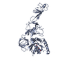
| Entry | Database: PDB / ID: 5djo | ||||||
|---|---|---|---|---|---|---|---|
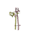
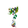
| Title | Crystal structure of the CC1-FHA tandem of Kinesin-3 KIF13A | ||||||
 Components Components | Kinesin-like protein | ||||||
 Keywords Keywords |  TRANSPORT PROTEIN / coiled-coil / FHA domain TRANSPORT PROTEIN / coiled-coil / FHA domain | ||||||
| Function / homology |  Function and homology information Function and homology informationmicrotubule motor activity => GO:0003777 / vesicle cargo loading / plus-end-directed vesicle transport along microtubule / Golgi to plasma membrane protein transport / melanosome organization / kinesin complex / microtubule motor activity / endosome to lysosome transport / microtubule-based movement / trans-Golgi network membrane ...microtubule motor activity => GO:0003777 / vesicle cargo loading / plus-end-directed vesicle transport along microtubule / Golgi to plasma membrane protein transport / melanosome organization / kinesin complex / microtubule motor activity / endosome to lysosome transport / microtubule-based movement / trans-Golgi network membrane / regulation of cytokinesis / intracellular protein transport / midbody / microtubule binding / microtubule / endosome membrane / cell cycle / cell division / centrosome / ATP hydrolysis activity / ATP bindingSimilarity search - Function | ||||||
| Biological species |  Mus musculus (house mouse) Mus musculus (house mouse) | ||||||
| Method | X-RAY DIFFRACTION / SYNCHROTRON / MOLECULAR REPLACEMENT / Resolution: 1.74 Å | ||||||
 Authors Authors | Ren, J.Q. / Li, W. / Huo, L. / Feng, W. | ||||||
 Citation Citation | Journal: J.Biol.Chem. / Year: 2016 Title: Structural Correlation of the Neck Coil with the Coiled-coil (CC1)-Forkhead-associated (FHA) Tandem for Active Kinesin-3 KIF13A Authors: Ren, J. / Huo, L. / Wang, W. / Zhang, Y. / Li, W. / Lou, J. / Xu, T. / Feng, W. | ||||||
| History |
|
- Structure visualization
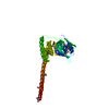
| Structure viewer | Molecule: MolmilJmol/JSmol |
|---|
- Downloads & links
-Download
| PDBx/mmCIF format | 5djo.cif.gz | 149.8 KB | Display | PDBx/mmCIF format |
|---|---|---|---|---|
| PDB format | pdb5djo.ent.gz | 117.1 KB | Display | PDB format |
| PDBx/mmJSON format | 5djo.json.gz | Tree view | PDBx/mmJSON format | |
| Others |  Other downloads Other downloads |
-Validation report
| Arichive directory | https://data.pdbj.org/pub/pdb/validation_reports/dj/5djoftp://data.pdbj.org/pub/pdb/validation_reports/dj/5djo | HTTPS FTP |
|---|
-Related structure data
| Related structure data | 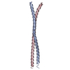 5djnC 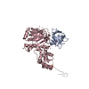 3fm8S C: citing same article ( S: Starting model for refinement |
|---|---|
| Similar structure data |


-Links
 PDBj
PDBj


- Assembly
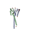
| Deposited unit | 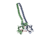
| ||||||||
|---|---|---|---|---|---|---|---|---|---|
| 1 |
| ||||||||
| Unit cell |
| ||||||||
| Components on special symmetry positions |
|
-Components
| #1: Protein | Mass: 19807.498 Da / Num. of mol.: 2 / Fragment: UNP residues 386-555 Source method: isolated from a genetically manipulated source Source: (gene. exp.) Mus musculus (house mouse) / Gene: Kif13a / Plasmid: pET32a / Production host:  Escherichia coli (E. coli) / References: UniProt: F8VQ75, UniProt: Q9EQW7*PLUS Escherichia coli (E. coli) / References: UniProt: F8VQ75, UniProt: Q9EQW7*PLUS#2: Chemical | ChemComp-FMT / Formic acid  Mass: 46.025 Da / Num. of mol.: 5 Mass: 46.025 Da / Num. of mol.: 5Source method: isolated from a genetically manipulated source Formula: CH2O2 #3: Chemical | ChemComp-ACY / | Acetic acid  Mass: 60.052 Da / Num. of mol.: 1 / Source method: obtained synthetically / Formula: C2H4O2 Mass: 60.052 Da / Num. of mol.: 1 / Source method: obtained synthetically / Formula: C2H4O2#4: Chemical | ChemComp-PEG / | Diethylene glycol  Mass: 106.120 Da / Num. of mol.: 1 / Source method: obtained synthetically / Formula: C4H10O3 Mass: 106.120 Da / Num. of mol.: 1 / Source method: obtained synthetically / Formula: C4H10O3#5: Water | ChemComp-HOH / | Water Mass: 18.015 Da / Num. of mol.: 264 / Source method: isolated from a natural source / Formula: H2O Mass: 18.015 Da / Num. of mol.: 264 / Source method: isolated from a natural source / Formula: H2O |
|---|
-Experimental details
-Experiment
| Experiment | Method: X-RAY DIFFRACTION / Number of used crystals: 1 |
|---|
- Sample preparation
| Crystal | Density Matthews: 2.09 Å3/Da / Density % sol: 41.25 % |
|---|---|
| Crystal grow | Temperature: 293 K / Method: evaporation / Details: 0.2 M calcium acetate, 16% (w/v) PEG 3350 |
-Data collection
| Diffraction | Mean temperature: 100 K |
|---|---|
| Diffraction source | Source: SYNCHROTRON / Site: SSRF  / Beamline: BL17U / Wavelength: 0.979 Å / Beamline: BL17U / Wavelength: 0.979 Å |
| Detector | Type: ADSC QUANTUM 315r / Detector: CCD / Date: Jul 12, 2012 |
| Radiation | Protocol: SINGLE WAVELENGTH / Monochromatic (M) / Laue (L): M / Scattering type: x-ray |
| Radiation wavelength | Wavelength: 0.979 Å / Relative weight: 1 |
| Reflection | Resolution: 1.74→49.55 Å / Num. obs: 33409 / % possible obs: 99 % / Redundancy: 5.1 % / Net I/σ(I): 12.2 |
| Reflection shell | Resolution: 1.74→1.79 Å / Redundancy: 5.3 % / Mean I/σ(I) obs: 2.6 / % possible all: 99.7 |
- Processing
| Software |
| ||||||||||||||||||||||||||||||||||||||||||||||||||||||||||||||||||||||||||||||||||||
|---|---|---|---|---|---|---|---|---|---|---|---|---|---|---|---|---|---|---|---|---|---|---|---|---|---|---|---|---|---|---|---|---|---|---|---|---|---|---|---|---|---|---|---|---|---|---|---|---|---|---|---|---|---|---|---|---|---|---|---|---|---|---|---|---|---|---|---|---|---|---|---|---|---|---|---|---|---|---|---|---|---|---|---|---|---|
| Refinement | Method to determine structure: MOLECULAR REPLACEMENT Starting model: 3FM8 Resolution: 1.74→40.488 Å / SU ML: 0.2 / Cross valid method: FREE R-VALUE / σ(F): 1.37 / Phase error: 21.32 / Stereochemistry target values: ML
| ||||||||||||||||||||||||||||||||||||||||||||||||||||||||||||||||||||||||||||||||||||
| Solvent computation | Shrinkage radii: 0.9 Å / VDW probe radii: 1.11 Å / Solvent model: FLAT BULK SOLVENT MODEL | ||||||||||||||||||||||||||||||||||||||||||||||||||||||||||||||||||||||||||||||||||||
| Displacement parameters | Biso max: 91.77 Å2 / Biso mean: 38.5082 Å2 / Biso min: 14.04 Å2 | ||||||||||||||||||||||||||||||||||||||||||||||||||||||||||||||||||||||||||||||||||||
| Refinement step | Cycle: final / Resolution: 1.74→40.488 Å
| ||||||||||||||||||||||||||||||||||||||||||||||||||||||||||||||||||||||||||||||||||||
| Refine LS restraints |
| ||||||||||||||||||||||||||||||||||||||||||||||||||||||||||||||||||||||||||||||||||||
| LS refinement shell |
|
