 Movie
Movie Controller


+ Open data

- Basic information
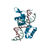
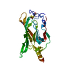
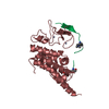
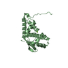
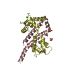
| Entry | Database: PDB / ID: 5dfn | ||||||
|---|---|---|---|---|---|---|---|
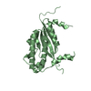
| Title | Structure of Tetrahymena Telomerase P45 C-terminal domain | ||||||
 Components Components | Telomerase associated protein p45 | ||||||
 Keywords Keywords |  NUCLEAR PROTEIN / Telomerase / P45 / CST complex / Stn1 / Winged Helix / WH domain / WHtH / Winged Helix turn Helix NUCLEAR PROTEIN / Telomerase / P45 / CST complex / Stn1 / Winged Helix / WH domain / WHtH / Winged Helix turn Helix | ||||||
| Function / homology | telomerase holoenzyme complex / telomere maintenance via telomerase / chromosome, telomeric region / metal ion binding / Telomerase-associated protein of 45 kDa / Telomerase-associated protein of 45 kDa Function and homology information Function and homology information | ||||||
| Biological species |   Tetrahymena thermophila (eukaryote) Tetrahymena thermophila (eukaryote) | ||||||
| Method | X-RAY DIFFRACTION / SYNCHROTRON / MOLECULAR REPLACEMENT / molecular replacement / Resolution: 2.382 Å | ||||||
| Model details | Telomerase protein P45C | ||||||
 Authors Authors | Chan, H. / Cascio, D. / Sawaya, M.R. / Feigon, J. | ||||||
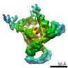
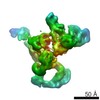
 Citation Citation | Journal: Science / Year: 2015 Title: Structure of Tetrahymena telomerase reveals previously unknown subunits, functions, and interactions. Authors: Jiansen Jiang / Henry Chan / Darian D Cash / Edward J Miracco / Rachel R Ogorzalek Loo / Heather E Upton / Duilio Cascio / Reid O'Brien Johnson / Kathleen Collins / Joseph A Loo / Z Hong Zhou / Juli Feigon /  Abstract: Telomerase helps maintain telomeres by processive synthesis of telomere repeat DNA at their 3'-ends, using an integral telomerase RNA (TER) and telomerase reverse transcriptase (TERT). We report the ...Telomerase helps maintain telomeres by processive synthesis of telomere repeat DNA at their 3'-ends, using an integral telomerase RNA (TER) and telomerase reverse transcriptase (TERT). We report the cryo-electron microscopy structure of Tetrahymena telomerase at ~9 angstrom resolution. In addition to seven known holoenzyme proteins, we identify two additional proteins that form a complex (TEB) with single-stranded telomere DNA-binding protein Teb1, paralogous to heterotrimeric replication protein A (RPA). The p75-p45-p19 subcomplex is identified as another RPA-related complex, CST (CTC1-STN1-TEN1). This study reveals the paths of TER in the TERT-TER-p65 catalytic core and single-stranded DNA exit; extensive subunit interactions of the TERT essential N-terminal domain, p50, and TEB; and other subunit identities and structures, including p19 and p45C crystal structures. Our findings provide structural and mechanistic insights into telomerase holoenzyme function. | ||||||
| History |
|
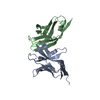
- Structure visualization
| Structure viewer | Molecule: MolmilJmol/JSmol |
|---|
- Downloads & links
-Download
| PDBx/mmCIF format | 5dfn.cif.gz | 149.2 KB | Display | PDBx/mmCIF format |
|---|---|---|---|---|
| PDB format | pdb5dfn.ent.gz | 117.3 KB | Display | PDB format |
| PDBx/mmJSON format | 5dfn.json.gz | Tree view | PDBx/mmJSON format | |
| Others |  Other downloads Other downloads |
-Validation report
| Arichive directory | https://data.pdbj.org/pub/pdb/validation_reports/df/5dfnftp://data.pdbj.org/pub/pdb/validation_reports/df/5dfn | HTTPS FTP |
|---|
-Related structure data


-Links
 PDBj
PDBj
- Assembly
| Deposited unit | 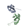
| |||||||||||||||||||||||||||||||||
|---|---|---|---|---|---|---|---|---|---|---|---|---|---|---|---|---|---|---|---|---|---|---|---|---|---|---|---|---|---|---|---|---|---|---|
| 1 | 
| |||||||||||||||||||||||||||||||||
| 2 | 
| |||||||||||||||||||||||||||||||||
| 3 | 
| |||||||||||||||||||||||||||||||||
| 4 | 
| |||||||||||||||||||||||||||||||||
| Unit cell |
| |||||||||||||||||||||||||||||||||
| Components on special symmetry positions |
| |||||||||||||||||||||||||||||||||
| Noncrystallographic symmetry (NCS) | NCS domain:
NCS domain segments: Component-ID: 1 / Ens-ID: 1 / Beg auth comp-ID: THR / Beg label comp-ID: THR
|
-Components
| #1: Protein | Mass: 25210.664 Da / Num. of mol.: 2 / Fragment: C-terminal domain (UNP residues 161-373) Source method: isolated from a genetically manipulated source Source: (gene. exp.) Tetrahymena thermophila (eukaryote) / Gene: P45 / Plasmid: pETDuet / Production host:  Escherichia coli (E. coli) / Strain (production host): BL21(DE3) / References: UniProt: Q237B3, UniProt: Q6JXI5*PLUS Escherichia coli (E. coli) / Strain (production host): BL21(DE3) / References: UniProt: Q237B3, UniProt: Q6JXI5*PLUS#2: Water | ChemComp-HOH / | Water Mass: 18.015 Da / Num. of mol.: 6 / Source method: isolated from a natural source / Formula: H2O Mass: 18.015 Da / Num. of mol.: 6 / Source method: isolated from a natural source / Formula: H2O |
|---|
-Experimental details
-Experiment
| Experiment | Method: X-RAY DIFFRACTION / Number of used crystals: 1 |
|---|
- Sample preparation
| Crystal | Density Matthews: 2.36 Å3/Da / Density % sol: 47.95 % |
|---|---|
| Crystal grow | Temperature: 298 K / Method: vapor diffusion, hanging drop / pH: 7.5 Details: 14 mg/ml P45 protein, 0.1 M Na-HEPES buffer, 5%v/v MPD, 10%w/v PEG-6000 |
-Data collection
| Diffraction | Mean temperature: 100 K | |||||||||||||||||||||||||||||||||||||||||||||||||||||||||||||||||||||||||||||||||||||||||||||||||||||||||||||||||||||||||||||||||||||||||||||||||||||||||||||||||||||||||||||||||||||||||||||
|---|---|---|---|---|---|---|---|---|---|---|---|---|---|---|---|---|---|---|---|---|---|---|---|---|---|---|---|---|---|---|---|---|---|---|---|---|---|---|---|---|---|---|---|---|---|---|---|---|---|---|---|---|---|---|---|---|---|---|---|---|---|---|---|---|---|---|---|---|---|---|---|---|---|---|---|---|---|---|---|---|---|---|---|---|---|---|---|---|---|---|---|---|---|---|---|---|---|---|---|---|---|---|---|---|---|---|---|---|---|---|---|---|---|---|---|---|---|---|---|---|---|---|---|---|---|---|---|---|---|---|---|---|---|---|---|---|---|---|---|---|---|---|---|---|---|---|---|---|---|---|---|---|---|---|---|---|---|---|---|---|---|---|---|---|---|---|---|---|---|---|---|---|---|---|---|---|---|---|---|---|---|---|---|---|---|---|---|---|---|---|
| Diffraction source | Source: SYNCHROTRON / Site: APS / Beamline: 24-ID-C / Wavelength: 0.9791 Å | |||||||||||||||||||||||||||||||||||||||||||||||||||||||||||||||||||||||||||||||||||||||||||||||||||||||||||||||||||||||||||||||||||||||||||||||||||||||||||||||||||||||||||||||||||||||||||||
| Detector | Type: DECTRIS PILATUS 6M / Detector: PIXEL / Date: Oct 16, 2014 | |||||||||||||||||||||||||||||||||||||||||||||||||||||||||||||||||||||||||||||||||||||||||||||||||||||||||||||||||||||||||||||||||||||||||||||||||||||||||||||||||||||||||||||||||||||||||||||
| Radiation | Protocol: SINGLE WAVELENGTH / Monochromatic (M) / Laue (L): M / Scattering type: x-ray | |||||||||||||||||||||||||||||||||||||||||||||||||||||||||||||||||||||||||||||||||||||||||||||||||||||||||||||||||||||||||||||||||||||||||||||||||||||||||||||||||||||||||||||||||||||||||||||
| Radiation wavelength | Wavelength: 0.9791 Å / Relative weight: 1 | |||||||||||||||||||||||||||||||||||||||||||||||||||||||||||||||||||||||||||||||||||||||||||||||||||||||||||||||||||||||||||||||||||||||||||||||||||||||||||||||||||||||||||||||||||||||||||||
| Reflection | Resolution: 2.38→86.89 Å / Num. obs: 18884 / % possible obs: 97.8 % / Observed criterion σ(I): -3 / Redundancy: 5.35 % / Biso Wilson estimate: 53.1 Å2 / Rmerge F obs: 0.999 / Rmerge(I) obs: 0.064 / Rrim(I) all: 0.072 / Χ2: 1.034 / Net I/σ(I): 14.07 / Num. measured all: 101012 | |||||||||||||||||||||||||||||||||||||||||||||||||||||||||||||||||||||||||||||||||||||||||||||||||||||||||||||||||||||||||||||||||||||||||||||||||||||||||||||||||||||||||||||||||||||||||||||
| Reflection shell | Diffraction-ID: 1 / Rejects: 0
|
-Phasing
| Phasing | Method: molecular replacement |
|---|
- Processing
| Software |
| ||||||||||||||||||||||||||||||||||||||||||||||||||||||||||||||||||||||||||||||||||||||||||||||||||
|---|---|---|---|---|---|---|---|---|---|---|---|---|---|---|---|---|---|---|---|---|---|---|---|---|---|---|---|---|---|---|---|---|---|---|---|---|---|---|---|---|---|---|---|---|---|---|---|---|---|---|---|---|---|---|---|---|---|---|---|---|---|---|---|---|---|---|---|---|---|---|---|---|---|---|---|---|---|---|---|---|---|---|---|---|---|---|---|---|---|---|---|---|---|---|---|---|---|---|---|
| Refinement | Method to determine structure: MOLECULAR REPLACEMENT / Resolution: 2.382→86.885 Å / SU ML: 0.37 / Cross valid method: FREE R-VALUE / σ(F): 1.36 / Phase error: 33.97 / Stereochemistry target values: ML
| ||||||||||||||||||||||||||||||||||||||||||||||||||||||||||||||||||||||||||||||||||||||||||||||||||
| Solvent computation | Shrinkage radii: 0.9 Å / VDW probe radii: 1.11 Å / Solvent model: FLAT BULK SOLVENT MODEL | ||||||||||||||||||||||||||||||||||||||||||||||||||||||||||||||||||||||||||||||||||||||||||||||||||
| Displacement parameters | Biso max: 161.49 Å2 / Biso mean: 62.762 Å2 / Biso min: 29.84 Å2 | ||||||||||||||||||||||||||||||||||||||||||||||||||||||||||||||||||||||||||||||||||||||||||||||||||
| Refinement step | Cycle: final / Resolution: 2.382→86.885 Å
| ||||||||||||||||||||||||||||||||||||||||||||||||||||||||||||||||||||||||||||||||||||||||||||||||||
| Refine LS restraints |
| ||||||||||||||||||||||||||||||||||||||||||||||||||||||||||||||||||||||||||||||||||||||||||||||||||
| Refine LS restraints NCS |
| ||||||||||||||||||||||||||||||||||||||||||||||||||||||||||||||||||||||||||||||||||||||||||||||||||
| LS refinement shell | Refine-ID: X-RAY DIFFRACTION / Total num. of bins used: 13
| ||||||||||||||||||||||||||||||||||||||||||||||||||||||||||||||||||||||||||||||||||||||||||||||||||
| Refinement TLS params. | Method: refined / Origin x: 76.4309 Å / Origin y: 15.5674 Å / Origin z: 148.336 Å
| ||||||||||||||||||||||||||||||||||||||||||||||||||||||||||||||||||||||||||||||||||||||||||||||||||
| Refinement TLS group |
|
