 Movie
Movie Controller

[English] 日本語
 Yorodumi
- PDB-5cx6: Crystal structure of Thosea asigna virus RNA-dependent RNA polyme... -
+ Open data

- Basic information
| Entry | Database: PDB / ID: 5cx6 | ||||||||||||
|---|---|---|---|---|---|---|---|---|---|---|---|---|---|
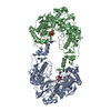
| Title | Crystal structure of Thosea asigna virus RNA-dependent RNA polymerase (RdRP) complexed with CDP | ||||||||||||
 Components Components | RNA-dependent RNA polymerase | ||||||||||||
 Keywords Keywords | TRANSFERASE / RdRP / complex / CDP | ||||||||||||
| Function / homology | RNA-dependent RNA polymerase activity / DNA/RNA polymerase superfamily / ATP binding / CYTIDINE-5'-DIPHOSPHATE / RNA-dependent RNA polymerase Function and homology information Function and homology information | ||||||||||||
| Biological species |  Thosea asigna virus Thosea asigna virus | ||||||||||||
| Method | X-RAY DIFFRACTION / SYNCHROTRON / MOLECULAR REPLACEMENT / Resolution: 2.1 Å | ||||||||||||
 Authors Authors | Ferrero, D.S. / Buxaderas, M. / Rodriguez, J.F. / Verdaguer, N. | ||||||||||||
| Funding support |  Spain, 3items Spain, 3items
| ||||||||||||
 Citation Citation | Journal: Plos Pathog. / Year: 2015 Title: The Structure of the RNA-Dependent RNA Polymerase of a Permutotetravirus Suggests a Link between Primer-Dependent and Primer-Independent Polymerases. Authors: Ferrero, D.S. / Buxaderas, M. / Rodriguez, J.F. / Verdaguer, N. | ||||||||||||
| History |
|
- Structure visualization
| Structure viewer | Molecule: MolmilJmol/JSmol |
|---|
- Downloads & links
-Download
| PDBx/mmCIF format | 5cx6.cif.gz | 558.4 KB | Display | PDBx/mmCIF format |
|---|---|---|---|---|
| PDB format | pdb5cx6.ent.gz | 455.3 KB | Display | PDB format |
| PDBx/mmJSON format | 5cx6.json.gz | Tree view | PDBx/mmJSON format | |
| Others |  Other downloads Other downloads |
-Validation report
| Arichive directory | https://data.pdbj.org/pub/pdb/validation_reports/cx/5cx6ftp://data.pdbj.org/pub/pdb/validation_reports/cx/5cx6 | HTTPS FTP |
|---|
-Related structure data
| Related structure data |  4xhaC 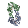 4xhiSC  5cyrC S: Starting model for refinement C: citing same article ( |
|---|---|
| Similar structure data |


-Links
 PDBj
PDBj- Assembly
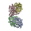
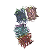
| Deposited unit | 
| ||||||||
|---|---|---|---|---|---|---|---|---|---|
| 1 |
| ||||||||
| Unit cell |
|
-Components
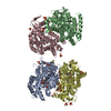
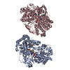
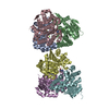
-Protein , 1 types, 2 molecules AB
| #1: Protein | Mass: 79540.461 Da / Num. of mol.: 2 / Fragment: UNP residues 1-674 Source method: isolated from a genetically manipulated source Source: (gene. exp.) Thosea asigna virus / Gene: RdRp / Plasmid: pFastBac1-HM / Production host:  Trichoplusia ni (cabbage looper) / References: UniProt: Q6A562 Trichoplusia ni (cabbage looper) / References: UniProt: Q6A562 |
|---|
-Non-polymers , 5 types, 637 molecules 


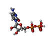

| #2: Chemical | ChemComp-SO4 / Sulfate Mass: 96.063 Da / Num. of mol.: 12 / Source method: obtained synthetically / Formula: SO4 Mass: 96.063 Da / Num. of mol.: 12 / Source method: obtained synthetically / Formula: SO4#3: Chemical | ChemComp-GOL / Glycerol Mass: 92.094 Da / Num. of mol.: 19 / Source method: obtained synthetically / Formula: C3H8O3 Mass: 92.094 Da / Num. of mol.: 19 / Source method: obtained synthetically / Formula: C3H8O3#4: Chemical | ChemComp-MG / |  Mass: 24.305 Da / Num. of mol.: 1 / Source method: obtained synthetically / Formula: Mg Mass: 24.305 Da / Num. of mol.: 1 / Source method: obtained synthetically / Formula: Mg#5: Chemical | Cytidine diphosphate Mass: 403.176 Da / Num. of mol.: 2 / Source method: obtained synthetically / Formula: C9H15N3O11P2 Mass: 403.176 Da / Num. of mol.: 2 / Source method: obtained synthetically / Formula: C9H15N3O11P2#6: Water | ChemComp-HOH / | WaterMass: 18.015 Da / Num. of mol.: 603 / Source method: isolated from a natural source / Formula: H2O |
|---|
-Experimental details
-Experiment
| Experiment | Method: X-RAY DIFFRACTION |
|---|
- Sample preparation
| Crystal | Density Matthews: 3.16 Å3/Da / Density % sol: 61.05 % |
|---|---|
| Crystal grow | Temperature: 293.15 K / Method: vapor diffusion, sitting drop / Details: PEG 8000, Lithium sulfate |
-Data collection
| Diffraction | Mean temperature: 80 K |
|---|---|
| Diffraction source | Source: SYNCHROTRON / Site: ESRF  / Beamline: ID29 / Wavelength: 0.976 Å / Beamline: ID29 / Wavelength: 0.976 Å |
| Detector | Type: ADSC QUANTUM 315r / Detector: CCD / Date: Apr 1, 2011 |
| Radiation | Protocol: SINGLE WAVELENGTH / Monochromatic (M) / Laue (L): M / Scattering type: x-ray |
| Radiation wavelength | Wavelength: 0.976 Å / Relative weight: 1 |
| Reflection | Resolution: 2.1→66.85 Å / Num. obs: 115494 / % possible obs: 98.31 % / Redundancy: 3.1 % / Rmerge(I) obs: 0.115 / Rsym value: 0.115 / Net I/σ(I): 5.64 |
| Reflection shell | Resolution: 2.1→2.175 Å / Redundancy: 3 % / Rmerge(I) obs: 0.422 / Mean I/σ(I) obs: 1.8 / % possible all: 98.6 |
- Processing
| Software |
| |||||||||||||||||||||||||||||||||||||||||||||||||||||||||||||||||||||||||||||||||||||||||||||||||||||||||||||||||||||||||||||||||||||||||||||||||||||||||||||||||||||||||||||||||||||||||||||||||||||||||||||||||||||||||
|---|---|---|---|---|---|---|---|---|---|---|---|---|---|---|---|---|---|---|---|---|---|---|---|---|---|---|---|---|---|---|---|---|---|---|---|---|---|---|---|---|---|---|---|---|---|---|---|---|---|---|---|---|---|---|---|---|---|---|---|---|---|---|---|---|---|---|---|---|---|---|---|---|---|---|---|---|---|---|---|---|---|---|---|---|---|---|---|---|---|---|---|---|---|---|---|---|---|---|---|---|---|---|---|---|---|---|---|---|---|---|---|---|---|---|---|---|---|---|---|---|---|---|---|---|---|---|---|---|---|---|---|---|---|---|---|---|---|---|---|---|---|---|---|---|---|---|---|---|---|---|---|---|---|---|---|---|---|---|---|---|---|---|---|---|---|---|---|---|---|---|---|---|---|---|---|---|---|---|---|---|---|---|---|---|---|---|---|---|---|---|---|---|---|---|---|---|---|---|---|---|---|---|---|---|---|---|---|---|---|---|---|---|---|---|---|---|---|---|
| Refinement | Method to determine structure: MOLECULAR REPLACEMENT Starting model: 4XHI Resolution: 2.1→66.85 Å / SU ML: 0.25 / Cross valid method: FREE R-VALUE / σ(F): 1.34 / Phase error: 22.06 / Stereochemistry target values: ML
| |||||||||||||||||||||||||||||||||||||||||||||||||||||||||||||||||||||||||||||||||||||||||||||||||||||||||||||||||||||||||||||||||||||||||||||||||||||||||||||||||||||||||||||||||||||||||||||||||||||||||||||||||||||||||
| Solvent computation | Shrinkage radii: 0.9 Å / VDW probe radii: 1.11 Å / Solvent model: FLAT BULK SOLVENT MODEL | |||||||||||||||||||||||||||||||||||||||||||||||||||||||||||||||||||||||||||||||||||||||||||||||||||||||||||||||||||||||||||||||||||||||||||||||||||||||||||||||||||||||||||||||||||||||||||||||||||||||||||||||||||||||||
| Refinement step | Cycle: LAST / Resolution: 2.1→66.85 Å
| |||||||||||||||||||||||||||||||||||||||||||||||||||||||||||||||||||||||||||||||||||||||||||||||||||||||||||||||||||||||||||||||||||||||||||||||||||||||||||||||||||||||||||||||||||||||||||||||||||||||||||||||||||||||||
| Refine LS restraints |
| |||||||||||||||||||||||||||||||||||||||||||||||||||||||||||||||||||||||||||||||||||||||||||||||||||||||||||||||||||||||||||||||||||||||||||||||||||||||||||||||||||||||||||||||||||||||||||||||||||||||||||||||||||||||||
| LS refinement shell |
| |||||||||||||||||||||||||||||||||||||||||||||||||||||||||||||||||||||||||||||||||||||||||||||||||||||||||||||||||||||||||||||||||||||||||||||||||||||||||||||||||||||||||||||||||||||||||||||||||||||||||||||||||||||||||
| Refinement TLS params. | Method: refined / Origin x: 24.6923 Å / Origin y: 31.1448 Å / Origin z: 23.2677 Å
| |||||||||||||||||||||||||||||||||||||||||||||||||||||||||||||||||||||||||||||||||||||||||||||||||||||||||||||||||||||||||||||||||||||||||||||||||||||||||||||||||||||||||||||||||||||||||||||||||||||||||||||||||||||||||
| Refinement TLS group | Selection details: all |
