 Movie
Movie Controller

[English] 日本語
 Yorodumi
- PDB-2z6x: Crystal structure of 22G, the wild-type protein of the photoswitc... -
+ Open data

- Basic information
| Entry | Database: PDB / ID: 2z6x | |||||||||
|---|---|---|---|---|---|---|---|---|---|---|
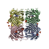
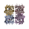
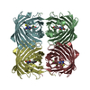
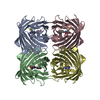
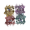
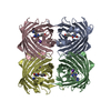
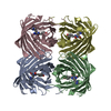
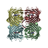
| Title | Crystal structure of 22G, the wild-type protein of the photoswitchable GFP-like protein Dronpa | |||||||||
 Components Components | photochromic protein Dronpa Photochromism Photochromism | |||||||||
 Keywords Keywords | FLUORESCENT PROTEIN / GFP-like Protein / Dronpa | |||||||||
| Function / homology |  Function and homology informationbioluminescence / generation of precursor metabolites and energy / identical protein binding / metal ion binding Function and homology informationbioluminescence / generation of precursor metabolites and energy / identical protein binding / metal ion bindingSimilarity search - Function | |||||||||
| Biological species |  Pectiniidae (invertebrata) Pectiniidae (invertebrata) | |||||||||
| Method | X-RAY DIFFRACTION / SYNCHROTRON / MOLECULAR REPLACEMENT / Resolution: 2.3 Å | |||||||||
 Authors Authors | Kikuchi, A. / Jeyakanthan, J. / Taka, J. / Shiro, Y. / Mizuno, H. / Miyawaki, A. | |||||||||
 Citation Citation | Journal: Proc.Natl.Acad.Sci.Usa / Year: 2008 Title: Light-dependent regulation of structural flexibility in a photochromic fluorescent protein. Authors: Mizuno, H. / Mal, T.K. / Walchli, M. / Kikuchi, A. / Fukano, T. / Ando, R. / Jeyakanthan, J. / Taka, J. / Shiro, Y. / Ikura, M. / Miyawaki, A. | |||||||||
| History |
|
- Structure visualization
| Structure viewer | Molecule: MolmilJmol/JSmol |
|---|
- Downloads & links
-Download
| PDBx/mmCIF format | 2z6x.cif.gz | 371.8 KB | Display | PDBx/mmCIF format |
|---|---|---|---|---|
| PDB format | pdb2z6x.ent.gz | 303 KB | Display | PDB format |
| PDBx/mmJSON format | 2z6x.json.gz | Tree view | PDBx/mmJSON format | |
| Others |  Other downloads Other downloads |
-Validation report
| Arichive directory | https://data.pdbj.org/pub/pdb/validation_reports/z6/2z6xftp://data.pdbj.org/pub/pdb/validation_reports/z6/2z6x | HTTPS FTP |
|---|
-Related structure data
| Related structure data | 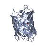 2z1oC 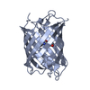 2z6yC 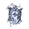 2z6zC 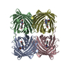 1ggxS C: citing same article ( S: Starting model for refinement |
|---|---|
| Similar structure data |


-Links
 PDBj
PDBj

- Assembly
| Deposited unit | 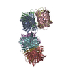
| ||||||||
|---|---|---|---|---|---|---|---|---|---|
| 1 | 
| ||||||||
| 2 | 
| ||||||||
| Unit cell |
|
-Components
| #1: Protein | Photochromism / GFP-like protein 22G / Fluorescent protein Dronpa Mass: 29230.107 Da / Num. of mol.: 8 Source method: isolated from a genetically manipulated source Source: (gene. exp.) Pectiniidae (invertebrata) / Plasmid: pRSET / Production host:  Escherichia coli (E. coli) / Strain (production host): JM109(DE3) / References: UniProt: Q5TLG6*PLUS Escherichia coli (E. coli) / Strain (production host): JM109(DE3) / References: UniProt: Q5TLG6*PLUS#2: Water | ChemComp-HOH / | Water Mass: 18.015 Da / Num. of mol.: 1081 / Source method: isolated from a natural source / Formula: H2O Mass: 18.015 Da / Num. of mol.: 1081 / Source method: isolated from a natural source / Formula: H2OSequence details | A SEQUENCE DATABASE REFERENCE FOR THIS PROTEIN DOES NOT CURRENTLY EXIST. THE RESIDUES (-32)-0 ARE ...A SEQUENCE DATABASE REFERENCE FOR THIS PROTEIN DOES NOT CURRENTLY EXIST. THE RESIDUES (-32)-0 ARE EXPRESSION | |
|---|
-Experimental details
-Experiment
| Experiment | Method: X-RAY DIFFRACTION / Number of used crystals: 1 |
|---|
- Sample preparation
| Crystal | Density Matthews: 2.09 Å3/Da / Density % sol: 41.07 % |
|---|---|
| Crystal grow | Temperature: 277 K / Method: vapor diffusion, sitting drop / pH: 8.5 Details: 0.2M Ammonium dihydrogen phosphate, 40 % MPD, 0.1M Tris-HCl (pH 8.5), VAPOR DIFFUSION, SITTING DROP, temperature 277K |
-Data collection
| Diffraction | Mean temperature: 100 K |
|---|---|
| Diffraction source | Source: SYNCHROTRON / Site: SPring-8  / Beamline: BL26B1 / Wavelength: 1 Å / Beamline: BL26B1 / Wavelength: 1 Å |
| Detector | Type: RIGAKU JUPITER 210 / Detector: CCD / Date: May 19, 2005 |
| Radiation | Monochromator: Si 111 / Protocol: SINGLE WAVELENGTH / Monochromatic (M) / Laue (L): M / Scattering type: x-ray |
| Radiation wavelength | Wavelength: 1 Å / Relative weight: 1 |
| Reflection | Resolution: 2.3→50 Å / Num. all: 85028 / Num. obs: 85028 / % possible obs: 100 % / Observed criterion σ(F): 0 / Observed criterion σ(I): 0 / Biso Wilson estimate: 22.2 Å2 / Rmerge(I) obs: 0.096 |
| Reflection shell | Resolution: 2.3→2.38 Å / Rmerge(I) obs: 0.361 / Mean I/σ(I) obs: 5.3 / Num. unique all: 8470 / % possible all: 100 |
- Processing
| Software |
| |||||||||||||||||||||||||
|---|---|---|---|---|---|---|---|---|---|---|---|---|---|---|---|---|---|---|---|---|---|---|---|---|---|---|
| Refinement | Method to determine structure: MOLECULAR REPLACEMENT Starting model: 1GGX Resolution: 2.3→19.96 Å / Rfactor Rfree error: 0.004 / Data cutoff high absF: 327000.99 / Data cutoff low absF: 0 / Isotropic thermal model: RESTRAINED / Cross valid method: THROUGHOUT / σ(F): 0 / Stereochemistry target values: Engh & Huber
| |||||||||||||||||||||||||
| Solvent computation | Solvent model: FLAT MODEL / Bsol: 37.1246 Å2 / ksol: 0.28926 e/Å3 | |||||||||||||||||||||||||
| Displacement parameters | Biso mean: 25.3 Å2
| |||||||||||||||||||||||||
| Refine analyze |
| |||||||||||||||||||||||||
| Refinement step | Cycle: LAST / Resolution: 2.3→19.96 Å
| |||||||||||||||||||||||||
| Refine LS restraints |
| |||||||||||||||||||||||||
| LS refinement shell | Resolution: 2.3→2.44 Å / Rfactor Rfree error: 0.013 / Total num. of bins used: 6
| |||||||||||||||||||||||||
| Xplor file |
|
