 Movie
Movie Controller

[English] 日本語
 Yorodumi
- PDB-5cwa: Structure of Anthranilate Synthase Component I (TrpE) from Mycoba... -
+ Open data

- Basic information
| Entry | Database: PDB / ID: 5cwa | |||||||||
|---|---|---|---|---|---|---|---|---|---|---|
| Title | Structure of Anthranilate Synthase Component I (TrpE) from Mycobacterium tuberculosis with inhibitor bound | |||||||||
 Components Components | Anthranilate synthase component 1 | |||||||||
 Keywords Keywords | Lyase/Lyase Inhibitor / Lyase / Inhibitor / Lyase-Lyase Inhibitor complex | |||||||||
| Function / homology |  Function and homology informationanthranilate synthase / anthranilate synthase activity / tryptophan biosynthetic process / metal ion binding Function and homology informationanthranilate synthase / anthranilate synthase activity / tryptophan biosynthetic process / metal ion bindingSimilarity search - Function | |||||||||
| Biological species |   Mycobacterium tuberculosis (bacteria) Mycobacterium tuberculosis (bacteria) | |||||||||
| Method | X-RAY DIFFRACTION / SYNCHROTRON / MOLECULAR REPLACEMENT / Resolution: 2.1 Å | |||||||||
 Authors Authors | Johnston, J.M. / Bashiri, G. / Evans, G.L. / Lott, J.S. / Baker, E.N. | |||||||||
 Citation Citation | Journal: Acta Crystallogr.,Sect.D / Year: 2015 Title: Structure and inhibition of subunit I of the anthranilate synthase complex of Mycobacterium tuberculosis and expression of the active complex. Authors: Bashiri, G. / Johnston, J.M. / Evans, G.L. / Bulloch, E.M. / Goldstone, D.C. / Jirgis, E.N. / Kleinboelting, S. / Castell, A. / Ramsay, R.J. / Manos-Turvey, A. / Payne, R.J. / Lott, J.S. / Baker, E.N. | |||||||||
| History |
|
- Structure visualization
| Structure viewer | Molecule: MolmilJmol/JSmol |
|---|
- Downloads & links
-Download
| PDBx/mmCIF format | 5cwa.cif.gz | 118.7 KB | Display | PDBx/mmCIF format |
|---|---|---|---|---|
| PDB format | pdb5cwa.ent.gz | 89.8 KB | Display | PDB format |
| PDBx/mmJSON format | 5cwa.json.gz | Tree view | PDBx/mmJSON format | |
| Others |  Other downloads Other downloads |
-Validation report
| Arichive directory | https://data.pdbj.org/pub/pdb/validation_reports/cw/5cwaftp://data.pdbj.org/pub/pdb/validation_reports/cw/5cwa | HTTPS FTP |
|---|
-Related structure data
| Related structure data | 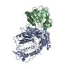 1qdlS S: Starting model for refinement |
|---|---|
| Similar structure data |


-Links
 PDBj
PDBj

- Assembly
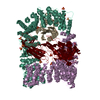
| Deposited unit | 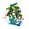
| ||||||||
|---|---|---|---|---|---|---|---|---|---|
| 1 | 
| ||||||||
| Unit cell |
| ||||||||
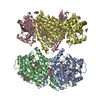
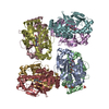
| Details | Biological assembly of TrpE is a dimer. In the structure this dimer is generated by application of crystal symmetry. This is supported by PISA analysis. Also further supported by experimental work - SEC and SEC-MALS analysis. |
-Components
-Protein , 1 types, 1 molecules A
| #1: Protein | / ASI Mass: 55496.258 Da / Num. of mol.: 1 / Mutation: H2D Source method: isolated from a genetically manipulated source Details: D TO H MUTATION FROM PUBLISHED SEQUENCE AT POSITION 2 DUE TO CLONING ARTEFACT, ALSO AFTER RTEV CLEAVAGE AN ADDITIONAL GA REMAINS ON CONSTRUCT USED PRIOR TO START M (NOT SHOWN IN SEQUENCE ABOVE) Source: (gene. exp.) Mycobacterium tuberculosis (strain CDC 1551 / Oshkosh) (bacteria)Strain: CDC 1551 / Oshkosh / Gene: trpE, MT1644 / Production host: Mycobacterium smegmatis (bacteria) / References: UniProt: P9WFX2, anthranilate synthase |
|---|
-Non-polymers , 5 types, 211 molecules 


| #2: Chemical | Sulfate Mass: 96.063 Da / Num. of mol.: 2 / Source method: obtained synthetically / Formula: SO4 Mass: 96.063 Da / Num. of mol.: 2 / Source method: obtained synthetically / Formula: SO4#3: Chemical | Glycerol Mass: 92.094 Da / Num. of mol.: 2 / Source method: obtained synthetically / Formula: C3H8O3 Mass: 92.094 Da / Num. of mol.: 2 / Source method: obtained synthetically / Formula: C3H8O3#4: Chemical | ChemComp-0GA / |  Mass: 238.193 Da / Num. of mol.: 1 / Source method: obtained synthetically / Formula: C11H10O6 Mass: 238.193 Da / Num. of mol.: 1 / Source method: obtained synthetically / Formula: C11H10O6#5: Chemical | ChemComp-UNL / | Num. of mol.: 1 / Source method: obtained synthetically #6: Water | ChemComp-HOH / | WaterMass: 18.015 Da / Num. of mol.: 205 / Source method: isolated from a natural source / Formula: H2O |
|---|
-Experimental details
-Experiment
| Experiment | Method: X-RAY DIFFRACTION / Number of used crystals: 1 |
|---|
- Sample preparation
| Crystal | Density Matthews: 4.09 Å3/Da / Density % sol: 69.95 % |
|---|---|
| Crystal grow | Temperature: 291 K / Method: vapor diffusion, hanging drop / pH: 7 Details: 0.6-1.0 M AMMONIUM SULFATE, 0.1 M BIS -TRIS PROPANE, PH 7, "silver bullet" additive G6, VAPOR DIFFUSION, TEMPERATURE 291K PH range: 7 |
-Data collection
| Diffraction | Mean temperature: 110 K |
|---|---|
| Diffraction source | Source: SYNCHROTRON / Site: Australian Synchrotron  / Beamline: MX2 / Wavelength: 0.8983 Å / Beamline: MX2 / Wavelength: 0.8983 Å |
| Detector | Type: ADSC QUANTUM 315r / Detector: CCD / Date: Mar 31, 2012 |
| Radiation | Protocol: SINGLE WAVELENGTH / Monochromatic (M) / Laue (L): M / Scattering type: x-ray |
| Radiation wavelength | Wavelength: 0.8983 Å / Relative weight: 1 |
| Reflection | Resolution: 2.1→135.452 Å / Num. obs: 54312 / % possible obs: 99.9 % / Redundancy: 21.9 % / Rsym value: 0.158 / Net I/σ(I): 14.8 |
| Reflection shell | Resolution: 2.1→2.21 Å / Redundancy: 22.4 % / Rmerge(I) obs: 3.794 / Mean I/σ(I) obs: 1.1 / Rsym value: 3.794 / % possible all: 99.8 |
- Processing
| Software |
| ||||||||||||||||||||||||||||||||||||||||||||||||||||||||||||||||||||||||||||||||||||||||||||||||||||||||||||||||||||||||||||||||||||||||||||||||||||||||||||||||||||||||||||||||||||||
|---|---|---|---|---|---|---|---|---|---|---|---|---|---|---|---|---|---|---|---|---|---|---|---|---|---|---|---|---|---|---|---|---|---|---|---|---|---|---|---|---|---|---|---|---|---|---|---|---|---|---|---|---|---|---|---|---|---|---|---|---|---|---|---|---|---|---|---|---|---|---|---|---|---|---|---|---|---|---|---|---|---|---|---|---|---|---|---|---|---|---|---|---|---|---|---|---|---|---|---|---|---|---|---|---|---|---|---|---|---|---|---|---|---|---|---|---|---|---|---|---|---|---|---|---|---|---|---|---|---|---|---|---|---|---|---|---|---|---|---|---|---|---|---|---|---|---|---|---|---|---|---|---|---|---|---|---|---|---|---|---|---|---|---|---|---|---|---|---|---|---|---|---|---|---|---|---|---|---|---|---|---|---|---|
| Refinement | Method to determine structure: MOLECULAR REPLACEMENT Starting model: 1QDL Resolution: 2.1→135.45 Å / Cor.coef. Fo:Fc: 0.963 / Cor.coef. Fo:Fc free: 0.942 / SU B: 5.884 / SU ML: 0.143 / Cross valid method: THROUGHOUT / σ(F): 0 / ESU R: 0.152 / ESU R Free: 0.153 / Stereochemistry target values: MAXIMUM LIKELIHOOD / Details: HYDROGENS HAVE BEEN ADDED IN THE RIDING
| ||||||||||||||||||||||||||||||||||||||||||||||||||||||||||||||||||||||||||||||||||||||||||||||||||||||||||||||||||||||||||||||||||||||||||||||||||||||||||||||||||||||||||||||||||||||
| Solvent computation | Ion probe radii: 0.8 Å / Shrinkage radii: 0.8 Å / VDW probe radii: 1.2 Å / Solvent model: MASK | ||||||||||||||||||||||||||||||||||||||||||||||||||||||||||||||||||||||||||||||||||||||||||||||||||||||||||||||||||||||||||||||||||||||||||||||||||||||||||||||||||||||||||||||||||||||
| Displacement parameters | Biso mean: 56.53 Å2
| ||||||||||||||||||||||||||||||||||||||||||||||||||||||||||||||||||||||||||||||||||||||||||||||||||||||||||||||||||||||||||||||||||||||||||||||||||||||||||||||||||||||||||||||||||||||
| Refinement step | Cycle: LAST / Resolution: 2.1→135.45 Å
| ||||||||||||||||||||||||||||||||||||||||||||||||||||||||||||||||||||||||||||||||||||||||||||||||||||||||||||||||||||||||||||||||||||||||||||||||||||||||||||||||||||||||||||||||||||||
| Refine LS restraints |
|