 Movie
Movie Controller

[English] 日本語
 Yorodumi
- PDB-1qdl: THE CRYSTAL STRUCTURE OF ANTHRANILATE SYNTHASE FROM SULFOLOBUS SO... -
+ Open data

- Basic information
| Entry | Database: PDB / ID: 1qdl | ||||||
|---|---|---|---|---|---|---|---|
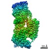
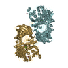
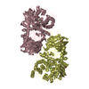
| Title | THE CRYSTAL STRUCTURE OF ANTHRANILATE SYNTHASE FROM SULFOLOBUS SOLFATARICUS | ||||||
 Components Components |
| ||||||
 Keywords Keywords |  LYASE / TRYPTOPHAN BIOSYNTHESIS / ANTHRANILATE SYNTHASE / GLUTAMINE AMIDOTRANSFERASE / ALLOSTERIC INTERACTION LYASE / TRYPTOPHAN BIOSYNTHESIS / ANTHRANILATE SYNTHASE / GLUTAMINE AMIDOTRANSFERASE / ALLOSTERIC INTERACTION | ||||||
| Function / homology |  Function and homology informationanthranilate synthase / anthranilate synthase activity / tryptophan biosynthetic process / glutamine metabolic process / metal ion binding Function and homology informationanthranilate synthase / anthranilate synthase activity / tryptophan biosynthetic process / glutamine metabolic process / metal ion bindingSimilarity search - Function | ||||||
| Biological species |   Sulfolobus solfataricus (archaea) Sulfolobus solfataricus (archaea) | ||||||
| Method | X-RAY DIFFRACTION / SYNCHROTRON / Resolution: 2.5 Å | ||||||
 Authors Authors | Knoechel, T. / Ivens, A. / Hester, G. / Gonzalez, A. / Bauerle, R. / Wilmanns, M. / Kirschner, K. / Jansonius, J.N. | ||||||
 Citation Citation | Journal: Proc.Natl.Acad.Sci.USA / Year: 1999 Title: The crystal structure of anthranilate synthase from Sulfolobus solfataricus: functional implications. Authors: Knochel, T. / Ivens, A. / Hester, G. / Gonzalez, A. / Bauerle, R. / Wilmanns, M. / Kirschner, K. / Jansonius, J.N. #1: Journal: Thesis, University Basel / Year: 1998Title: X-Ray Crystallographic Studies on Hyperthermostable Enzymes of the Tryptophan Biosynthesis Pathway: Three-Dimensional Structures of Indole-3-Glycerol Phosphate Synthase and Anthranilate Synthase Authors: Knoechel, T. | ||||||
| History |
|
- Structure visualization
| Structure viewer | Molecule: MolmilJmol/JSmol |
|---|
- Downloads & links
-Download
| PDBx/mmCIF format | 1qdl.cif.gz | 132.4 KB | Display | PDBx/mmCIF format |
|---|---|---|---|---|
| PDB format | pdb1qdl.ent.gz | 104.1 KB | Display | PDB format |
| PDBx/mmJSON format | 1qdl.json.gz | Tree view | PDBx/mmJSON format | |
| Others |  Other downloads Other downloads |
-Validation report
| Arichive directory | https://data.pdbj.org/pub/pdb/validation_reports/qd/1qdlftp://data.pdbj.org/pub/pdb/validation_reports/qd/1qdl | HTTPS FTP |
|---|
-Related structure data
| Similar structure data |
|---|


-Links
 PDBj
PDBj

- Assembly
| Deposited unit | 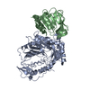
| ||||||||
|---|---|---|---|---|---|---|---|---|---|
| 1 | 
| ||||||||
| Unit cell |
|
-Components
| #1: Protein | Mass: 47779.453 Da / Num. of mol.: 1 / Fragment: AMINODEOXYISOCHORISMATE SYNTHASE/LYASE SUBUNIT Source method: isolated from a genetically manipulated source Details: CONTAINS RESIDUES GLY-3,SER-2, HIS-1 PRIOR TO THE INITIATING METHIONINE Source: (gene. exp.) Sulfolobus solfataricus (archaea) / Plasmid: PET15B / Production host:  Escherichia coli (E. coli) / References: UniProt: Q06128, anthranilate synthase Escherichia coli (E. coli) / References: UniProt: Q06128, anthranilate synthase |
|---|---|
| #2: Protein | Mass: 21937.258 Da / Num. of mol.: 1 / Fragment: GLUTAMINE AMIDOTRANSFERASE SUBUNIT Source method: isolated from a genetically manipulated source Source: (gene. exp.) Sulfolobus solfataricus (archaea) / Plasmid: PET15B / Production host: Escherichia coli (E. coli) / References: UniProt: Q06129, anthranilate synthase |
| #3: Water | ChemComp-HOH / Water Mass: 18.015 Da / Num. of mol.: 130 / Source method: isolated from a natural source / Formula: H2O Mass: 18.015 Da / Num. of mol.: 130 / Source method: isolated from a natural source / Formula: H2O |
-Experimental details
-Experiment
| Experiment | Method: X-RAY DIFFRACTION / Number of used crystals: 1 |
|---|
- Sample preparation
| Crystal | *PLUS Density % sol: 79 % | ||||||||||||||||||||||||||||||||||||||||||||||||
|---|---|---|---|---|---|---|---|---|---|---|---|---|---|---|---|---|---|---|---|---|---|---|---|---|---|---|---|---|---|---|---|---|---|---|---|---|---|---|---|---|---|---|---|---|---|---|---|---|---|
| Crystal grow | pH: 7.5 Details: SODIUM CHLORIDE, PEG 6000, POTASSIUM PHOSPHATE, pH 7.50 | ||||||||||||||||||||||||||||||||||||||||||||||||
| Crystal grow | *PLUS Method: vapor diffusionDetails: drop consists of equal volume of protein and reservoir solutions | ||||||||||||||||||||||||||||||||||||||||||||||||
| Components of the solutions | *PLUS
|
-Data collection
| Diffraction | Mean temperature: 100 K |
|---|---|
| Diffraction source | Source: SYNCHROTRON / Site: EMBL/DESY, HAMBURG  / Beamline: X11 / Wavelength: 0.9095 / Beamline: X11 / Wavelength: 0.9095 |
| Detector | Type: MARRESEARCH / Detector: IMAGE PLATE / Date: Jul 16, 1997 |
| Radiation | Protocol: SINGLE WAVELENGTH / Monochromatic (M) / Laue (L): M / Scattering type: x-ray |
| Radiation wavelength | Wavelength: 0.9095 Å / Relative weight: 1 |
| Reflection | Resolution: 2.5→30 Å / Num. obs: 57136 / % possible obs: 99.8 % / Observed criterion σ(I): -3 / Redundancy: 9.7 % / Biso Wilson estimate: 46.7 Å2 / Rmerge(I) obs: 0.054 / Net I/σ(I): 30.49 |
| Reflection shell | Resolution: 2.5→2.59 Å / Rmerge(I) obs: 0.291 / % possible all: 99.6 |
| Reflection shell | *PLUS % possible obs: 99.6 % |
- Processing
| Software |
| ||||||||||||||||||||||||||||||||||||||||||||||||||||||||||||
|---|---|---|---|---|---|---|---|---|---|---|---|---|---|---|---|---|---|---|---|---|---|---|---|---|---|---|---|---|---|---|---|---|---|---|---|---|---|---|---|---|---|---|---|---|---|---|---|---|---|---|---|---|---|---|---|---|---|---|---|---|---|
| Refinement | Resolution: 2.5→30 Å / σ(F): 1 Details: A FLAT BULK-SOLVENT MODEL WAS SUPPLIED THROUGHOUT THE REFINEMENT. DIFFRACTION DATA WERE SUBJECTED TO AN OVERALL ANISOTROPIC B-FACTOR SCALING.
| ||||||||||||||||||||||||||||||||||||||||||||||||||||||||||||
| Refinement step | Cycle: LAST / Resolution: 2.5→30 Å
| ||||||||||||||||||||||||||||||||||||||||||||||||||||||||||||
| Refine LS restraints |
|
