 Movie
Movie Controller


+ Open data

- Basic information
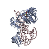
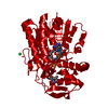
| Entry | Database: PDB / ID: 5cvl | ||||||
|---|---|---|---|---|---|---|---|
| Title | WDR48 (UAF-1), residues 2-580 | ||||||
 Components Components | WD repeat-containing protein 48 | ||||||
 Keywords Keywords |  PROTEIN BINDING / WDR48 / UAF1 / WD-repeat / USP / deubiquitinase / DUB PROTEIN BINDING / WDR48 / UAF1 / WD-repeat / USP / deubiquitinase / DUB | ||||||
| Function / homology |  Function and homology information Function and homology informationregulation of protein monoubiquitination / Signaling by cytosolic PDGFRA and PDGFRB fusion proteins / deubiquitinase activator activity / skeletal system morphogenesis / skin development / positive regulation of double-strand break repair via homologous recombination / seminiferous tubule development / single fertilization / homeostasis of number of cells / embryonic organ development ...regulation of protein monoubiquitination / Signaling by cytosolic PDGFRA and PDGFRB fusion proteins / deubiquitinase activator activity / skeletal system morphogenesis / skin development / positive regulation of double-strand break repair via homologous recombination / seminiferous tubule development / single fertilization / homeostasis of number of cells / embryonic organ development / ubiquitin binding / positive regulation of epithelial cell proliferation / Recognition of DNA damage by PCNA-containing replication complex / Fanconi Anemia Pathway / double-strand break repair via homologous recombination / positive regulation of receptor signaling pathway via JAK-STAT / multicellular organism growth / late endosome / single-stranded DNA binding / double-stranded DNA binding / spermatogenesis / lysosome / Ub-specific processing proteases / intracellular membrane-bounded organelle / DNA damage response / DNA binding / nucleoplasm / nucleus / cytosol / cytoplasmSimilarity search - Function | ||||||
| Biological species |  Homo sapiens (human) Homo sapiens (human) | ||||||
| Method | X-RAY DIFFRACTION / SYNCHROTRON / SAD / Resolution: 3 Å | ||||||
 Authors Authors | HARRIS, S.F. / YIN, J. | ||||||
 Citation Citation | Journal: Structure / Year: 2015 Title: Structural Insights into WD-Repeat 48 Activation of Ubiquitin-Specific Protease 46. Authors: Yin, J. / Schoeffler, A.J. / Wickliffe, K. / Newton, K. / Starovasnik, M.A. / Dueber, E.C. / Harris, S.F. | ||||||
| History |
|
- Structure visualization
| Structure viewer | Molecule: MolmilJmol/JSmol |
|---|
- Downloads & links
-Download
| PDBx/mmCIF format | 5cvl.cif.gz | 124 KB | Display | PDBx/mmCIF format |
|---|---|---|---|---|
| PDB format | pdb5cvl.ent.gz | 93.3 KB | Display | PDB format |
| PDBx/mmJSON format | 5cvl.json.gz | Tree view | PDBx/mmJSON format | |
| Others |  Other downloads Other downloads |
-Validation report
| Arichive directory | https://data.pdbj.org/pub/pdb/validation_reports/cv/5cvlftp://data.pdbj.org/pub/pdb/validation_reports/cv/5cvl | HTTPS FTP |
|---|
-Related structure data


-Links
 PDBj
PDBj


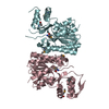
- Assembly
| Deposited unit | 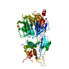
| ||||||||
|---|---|---|---|---|---|---|---|---|---|
| 1 |
| ||||||||
| Unit cell |
| ||||||||
| Components on special symmetry positions |
|
-Components
| #1: Protein | Mass: 67150.906 Da / Num. of mol.: 1 / Fragment: UNP residues 2-580 Source method: isolated from a genetically manipulated source Source: (gene. exp.) Homo sapiens (human) / Gene: WDR48, KIAA1449, UAF1 / Production host:  Trichoplusia ni (cabbage looper) / References: UniProt: Q8TAF3 Trichoplusia ni (cabbage looper) / References: UniProt: Q8TAF3 | ||||
|---|---|---|---|---|---|
| #2: Chemical | Phosphate  Mass: 94.971 Da / Num. of mol.: 2 / Source method: isolated from a natural source / Formula: PO4 Mass: 94.971 Da / Num. of mol.: 2 / Source method: isolated from a natural source / Formula: PO4#3: Chemical | ChemComp-AU /   Mass: 196.967 Da / Num. of mol.: 7 / Source method: obtained synthetically / Formula: Au Mass: 196.967 Da / Num. of mol.: 7 / Source method: obtained synthetically / Formula: Au#4: Water | ChemComp-HOH / | Water Mass: 18.015 Da / Num. of mol.: 137 / Source method: isolated from a natural source / Formula: H2O Mass: 18.015 Da / Num. of mol.: 137 / Source method: isolated from a natural source / Formula: H2O |
-Experimental details
-Experiment
| Experiment | Method: X-RAY DIFFRACTION / Number of used crystals: 1 |
|---|
- Sample preparation
| Crystal | Density Matthews: 4.26 Å3/Da / Density % sol: 71.12 % / Mosaicity: 0.359 ° |
|---|---|
| Crystal grow | Temperature: 292 K / Method: vapor diffusion, hanging drop / pH: 7.5 Details: 0.72 M sodium phosphate, 0.72 M potassium phosphate, 90 mM HEPES pH 7.5 |
-Data collection
| Diffraction | Mean temperature: 100 K | |||||||||||||||||||||||||||||||||||||||||||||||||||||||
|---|---|---|---|---|---|---|---|---|---|---|---|---|---|---|---|---|---|---|---|---|---|---|---|---|---|---|---|---|---|---|---|---|---|---|---|---|---|---|---|---|---|---|---|---|---|---|---|---|---|---|---|---|---|---|---|---|
| Diffraction source | Source: SYNCHROTRON / Site: SSRL  / Beamline: BL7-1 / Wavelength: 1.026382 Å / Beamline: BL7-1 / Wavelength: 1.026382 Å | |||||||||||||||||||||||||||||||||||||||||||||||||||||||
| Detector | Type: ADSC QUANTUM 315 / Detector: CCD / Date: Aug 1, 2012 | |||||||||||||||||||||||||||||||||||||||||||||||||||||||
| Radiation | Monochromator: DOUBLE CRYSTAL Si(111) / Protocol: SINGLE WAVELENGTH / Monochromatic (M) / Laue (L): M / Scattering type: x-ray | |||||||||||||||||||||||||||||||||||||||||||||||||||||||
| Radiation wavelength | Wavelength: 1.026382 Å / Relative weight: 1 | |||||||||||||||||||||||||||||||||||||||||||||||||||||||
| Reflection | Resolution: 3→50 Å / Num. obs: 44419 / % possible obs: 100 % / Redundancy: 8.6 % / Biso Wilson estimate: 81.5 Å2 / Rmerge(I) obs: 0.123 / Χ2: 1.069 / Net I/av σ(I): 19.429 / Net I/σ(I): 7.3 / Num. measured all: 381541 | |||||||||||||||||||||||||||||||||||||||||||||||||||||||
| Reflection shell | Diffraction-ID: 1 / Rejects: 0 / % possible all: 100
|
-Phasing
| Phasing | Method: SAD |
|---|
- Processing
| Software |
| ||||||||||||||||||||||||||||||||||||||||||||||||||||||||||||||||||||||||||||||||||||||||||||||||||||||||||||
|---|---|---|---|---|---|---|---|---|---|---|---|---|---|---|---|---|---|---|---|---|---|---|---|---|---|---|---|---|---|---|---|---|---|---|---|---|---|---|---|---|---|---|---|---|---|---|---|---|---|---|---|---|---|---|---|---|---|---|---|---|---|---|---|---|---|---|---|---|---|---|---|---|---|---|---|---|---|---|---|---|---|---|---|---|---|---|---|---|---|---|---|---|---|---|---|---|---|---|---|---|---|---|---|---|---|---|---|---|---|
| Refinement | Method to determine structure: SAD / Resolution: 3→49.33 Å / Cor.coef. Fo:Fc: 0.9324 / Cor.coef. Fo:Fc free: 0.8988 / SU R Cruickshank DPI: 0.455 / Cross valid method: THROUGHOUT / σ(F): 0 / SU R Blow DPI: 0.475 / SU Rfree Blow DPI: 0.297 / SU Rfree Cruickshank DPI: 0.297
| ||||||||||||||||||||||||||||||||||||||||||||||||||||||||||||||||||||||||||||||||||||||||||||||||||||||||||||
| Displacement parameters | Biso max: 175.41 Å2 / Biso mean: 69.95 Å2 / Biso min: 16.53 Å2
| ||||||||||||||||||||||||||||||||||||||||||||||||||||||||||||||||||||||||||||||||||||||||||||||||||||||||||||
| Refine analyze | Luzzati coordinate error obs: 0.378 Å | ||||||||||||||||||||||||||||||||||||||||||||||||||||||||||||||||||||||||||||||||||||||||||||||||||||||||||||
| Refinement step | Cycle: final / Resolution: 3→49.33 Å
| ||||||||||||||||||||||||||||||||||||||||||||||||||||||||||||||||||||||||||||||||||||||||||||||||||||||||||||
| Refine LS restraints |
| ||||||||||||||||||||||||||||||||||||||||||||||||||||||||||||||||||||||||||||||||||||||||||||||||||||||||||||
| LS refinement shell | Resolution: 3→3.13 Å / Total num. of bins used: 12
|
