 Movie
Movie Controller


+ Open data

- Basic information
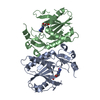
| Entry | Database: PDB / ID: 5l8e | ||||||||||||
|---|---|---|---|---|---|---|---|---|---|---|---|---|---|
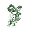
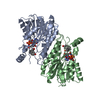
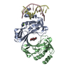
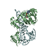
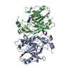
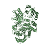
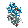
| Title | Structure of UAF1 | ||||||||||||
 Components Components |
| ||||||||||||
 Keywords Keywords |  STRUCTURAL PROTEIN / WDR48 Activates USP1/12/46 B propellar STRUCTURAL PROTEIN / WDR48 Activates USP1/12/46 B propellar | ||||||||||||
| Function / homology |  Function and homology information Function and homology informationregulation of protein monoubiquitination / Signaling by cytosolic PDGFRA and PDGFRB fusion proteins / deubiquitinase activator activity / skeletal system morphogenesis / skin development / seminiferous tubule development / positive regulation of double-strand break repair via homologous recombination / homeostasis of number of cells / single fertilization / embryonic organ development ...regulation of protein monoubiquitination / Signaling by cytosolic PDGFRA and PDGFRB fusion proteins / deubiquitinase activator activity / skeletal system morphogenesis / skin development / seminiferous tubule development / positive regulation of double-strand break repair via homologous recombination / homeostasis of number of cells / single fertilization / embryonic organ development / ubiquitin binding / positive regulation of epithelial cell proliferation / Fanconi Anemia Pathway / Recognition of DNA damage by PCNA-containing replication complex / double-strand break repair via homologous recombination / positive regulation of receptor signaling pathway via JAK-STAT / multicellular organism growth / late endosome / single-stranded DNA binding / spermatogenesis / double-stranded DNA binding / lysosome / Ub-specific processing proteases / intracellular membrane-bounded organelle / DNA damage response / DNA binding / nucleoplasm / nucleus / cytosol / cytoplasmSimilarity search - Function | ||||||||||||
| Biological species |  Homo sapiens (human) Homo sapiens (human) | ||||||||||||
| Method | X-RAY DIFFRACTION / SYNCHROTRON / MOLECULAR REPLACEMENT / Resolution: 2.3 Å | ||||||||||||
 Authors Authors | Dharadhar, S. / Sixma, T. | ||||||||||||
| Funding support |  Netherlands, 3items Netherlands, 3items
| ||||||||||||
 Citation Citation | Journal: J.Struct.Biol. / Year: 2016 Title: A conserved two-step binding for the UAF1 regulator to the USP12 deubiquitinating enzyme. Authors: Dharadhar, S. / Clerici, M. / van Dijk, W.J. / Fish, A. / Sixma, T.K. | ||||||||||||
| History |
|
- Structure visualization
| Structure viewer | Molecule: MolmilJmol/JSmol |
|---|
- Downloads & links
-Download
| PDBx/mmCIF format | 5l8e.cif.gz | 222 KB | Display | PDBx/mmCIF format |
|---|---|---|---|---|
| PDB format | pdb5l8e.ent.gz | 177.6 KB | Display | PDB format |
| PDBx/mmJSON format | 5l8e.json.gz | Tree view | PDBx/mmJSON format | |
| Others |  Other downloads Other downloads |
-Validation report
| Arichive directory | https://data.pdbj.org/pub/pdb/validation_reports/l8/5l8eftp://data.pdbj.org/pub/pdb/validation_reports/l8/5l8e | HTTPS FTP |
|---|
-Related structure data
| Related structure data | 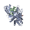 5l8hC  5l8wC  1vyhS S: Starting model for refinement C: citing same article ( |
|---|---|
| Similar structure data |


-Links
 PDBj
PDBj


- Assembly
| Deposited unit | 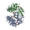
| ||||||||
|---|---|---|---|---|---|---|---|---|---|
| 1 | 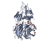
| ||||||||
| 2 | 
| ||||||||
| Unit cell |
|
-Components
| #1: Protein | Mass: 65194.832 Da / Num. of mol.: 2 Source method: isolated from a genetically manipulated source Source: (gene. exp.) Homo sapiens (human) / Gene: WDR48, KIAA1449, UAF1 / Production host:   Spodoptera frugiperda (fall armyworm) / References: UniProt: Q8TAF3 Spodoptera frugiperda (fall armyworm) / References: UniProt: Q8TAF3#2: Protein/peptide | | Mass: 443.539 Da / Num. of mol.: 1 Source method: isolated from a genetically manipulated source Source: (gene. exp.) Homo sapiens (human) / Production host: Spodoptera frugiperda (fall armyworm)#3: Chemical | ChemComp-GOL / Glycerol  Mass: 92.094 Da / Num. of mol.: 14 / Source method: obtained synthetically / Formula: C3H8O3 Mass: 92.094 Da / Num. of mol.: 14 / Source method: obtained synthetically / Formula: C3H8O3#4: Water | ChemComp-HOH / | Water Mass: 18.015 Da / Num. of mol.: 310 / Source method: isolated from a natural source / Formula: H2O Mass: 18.015 Da / Num. of mol.: 310 / Source method: isolated from a natural source / Formula: H2O |
|---|
-Experimental details
-Experiment
| Experiment | Method: X-RAY DIFFRACTION / Number of used crystals: 1 |
|---|
- Sample preparation
| Crystal | Density Matthews: 2.74 Å3/Da / Density % sol: 55.12 % |
|---|---|
| Crystal grow | Temperature: 293 K / Method: vapor diffusion, sitting drop / pH: 6.5 Details: 20% PEG3350, 200mM Tri-Sodium Citrate, Bis-Tris Propane Cryo -30% Glycerol |
-Data collection
| Diffraction | Mean temperature: 100 K |
|---|---|
| Diffraction source | Source: SYNCHROTRON / Site: SLS  / Beamline: X06DA / Wavelength: 0.91997 Å / Beamline: X06DA / Wavelength: 0.91997 Å |
| Detector | Type: DECTRIS PILATUS 2M / Detector: PIXEL / Date: Sep 4, 2015 |
| Radiation | Protocol: SINGLE WAVELENGTH / Monochromatic (M) / Laue (L): M / Scattering type: x-ray |
| Radiation wavelength | Wavelength: 0.91997 Å / Relative weight: 1 |
| Reflection | Resolution: 2.3→49 Å / Num. obs: 64580 / % possible obs: 99.6 % / Redundancy: 4.5 % / CC1/2: 0.999 / Rmerge(I) obs: 0.05 / Net I/σ(I): 16.1 |
| Reflection shell | Resolution: 2.3→2.35 Å / Redundancy: 4.1 % / Rmerge(I) obs: 1 / Mean I/σ(I) obs: 1.4 / % possible all: 95.9 |
- Processing
| Software |
| ||||||||||||||||||||||||||||||||||||||||||||||||||||||||||||||||||||||||||||||||||||||||||||||||||||||||||||||||||||||||||||||||||||||||||||||||||||||||||||||||||||||||||||||||||||||
|---|---|---|---|---|---|---|---|---|---|---|---|---|---|---|---|---|---|---|---|---|---|---|---|---|---|---|---|---|---|---|---|---|---|---|---|---|---|---|---|---|---|---|---|---|---|---|---|---|---|---|---|---|---|---|---|---|---|---|---|---|---|---|---|---|---|---|---|---|---|---|---|---|---|---|---|---|---|---|---|---|---|---|---|---|---|---|---|---|---|---|---|---|---|---|---|---|---|---|---|---|---|---|---|---|---|---|---|---|---|---|---|---|---|---|---|---|---|---|---|---|---|---|---|---|---|---|---|---|---|---|---|---|---|---|---|---|---|---|---|---|---|---|---|---|---|---|---|---|---|---|---|---|---|---|---|---|---|---|---|---|---|---|---|---|---|---|---|---|---|---|---|---|---|---|---|---|---|---|---|---|---|---|---|
| Refinement | Method to determine structure: MOLECULAR REPLACEMENT Starting model: 1VYH Resolution: 2.3→49 Å / Cor.coef. Fo:Fc: 0.965 / Cor.coef. Fo:Fc free: 0.95 / SU B: 7.24 / SU ML: 0.163 / Cross valid method: THROUGHOUT / ESU R: 0.245 / ESU R Free: 0.196 / Details: HYDROGENS HAVE BEEN ADDED IN THE RIDING POSITIONS
| ||||||||||||||||||||||||||||||||||||||||||||||||||||||||||||||||||||||||||||||||||||||||||||||||||||||||||||||||||||||||||||||||||||||||||||||||||||||||||||||||||||||||||||||||||||||
| Solvent computation | Ion probe radii: 0.8 Å / Shrinkage radii: 0.8 Å / VDW probe radii: 1.1 Å | ||||||||||||||||||||||||||||||||||||||||||||||||||||||||||||||||||||||||||||||||||||||||||||||||||||||||||||||||||||||||||||||||||||||||||||||||||||||||||||||||||||||||||||||||||||||
| Displacement parameters | Biso mean: 58.297 Å2
| ||||||||||||||||||||||||||||||||||||||||||||||||||||||||||||||||||||||||||||||||||||||||||||||||||||||||||||||||||||||||||||||||||||||||||||||||||||||||||||||||||||||||||||||||||||||
| Refinement step | Cycle: 1 / Resolution: 2.3→49 Å
| ||||||||||||||||||||||||||||||||||||||||||||||||||||||||||||||||||||||||||||||||||||||||||||||||||||||||||||||||||||||||||||||||||||||||||||||||||||||||||||||||||||||||||||||||||||||
| Refine LS restraints |
|