 Movie
Movie Controller


+ Open data

- Basic information
| Entry | Database: PDB / ID: 3aw9 | ||||||
|---|---|---|---|---|---|---|---|
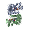
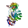
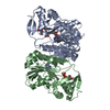
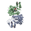
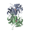
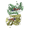
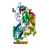
| Title | Structure of UDP-galactose 4-epimerase mutant | ||||||
 Components Components | NAD-dependent epimerase/dehydratase | ||||||
 Keywords Keywords |  ISOMERASE / Rossmann fold ISOMERASE / Rossmann fold | ||||||
| Function / homology |  Function and homology informationUDP-glucose 4-epimerase activity / galactose catabolic process via UDP-galactose / oxidoreductase activity / nucleotide binding / cytosol Function and homology informationUDP-glucose 4-epimerase activity / galactose catabolic process via UDP-galactose / oxidoreductase activity / nucleotide binding / cytosolSimilarity search - Function | ||||||
| Biological species |   Pyrobaculum calidifontis (archaea) Pyrobaculum calidifontis (archaea) | ||||||
| Method | X-RAY DIFFRACTION / SYNCHROTRON / MOLECULAR REPLACEMENT / molecular replacement / Resolution: 2.3 Å | ||||||
 Authors Authors | Sakuraba, H. / Kawai, T. / Yoneda, K. / Ohshima, T. | ||||||
 Citation Citation | Journal: To be Published Title: Structure of UDP-galactose 4-epimerase mutant Authors: Sakuraba, H. / Kawai, T. / Yoneda, K. / Ohshima, T. | ||||||
| History |
|
- Structure visualization
| Structure viewer | Molecule: MolmilJmol/JSmol |
|---|
- Downloads & links
-Download
| PDBx/mmCIF format | 3aw9.cif.gz | 191.9 KB | Display | PDBx/mmCIF format |
|---|---|---|---|---|
| PDB format | pdb3aw9.ent.gz | 158.4 KB | Display | PDB format |
| PDBx/mmJSON format | 3aw9.json.gz | Tree view | PDBx/mmJSON format | |
| Others |  Other downloads Other downloads |
-Validation report
| Arichive directory | https://data.pdbj.org/pub/pdb/validation_reports/aw/3aw9ftp://data.pdbj.org/pub/pdb/validation_reports/aw/3aw9 | HTTPS FTP |
|---|
-Related structure data
| Similar structure data |
|---|


-Links
 PDBj
PDBj


- Assembly
| Deposited unit | 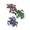
| ||||||||
|---|---|---|---|---|---|---|---|---|---|
| 1 | 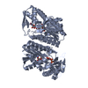
| ||||||||
| 2 | 
| ||||||||
| Unit cell |
|
-Components
| #1: Protein | Mass: 33864.934 Da / Num. of mol.: 3 / Mutation: DELETION MUTANT Source method: isolated from a genetically manipulated source Source: (gene. exp.) Pyrobaculum calidifontis (archaea) / Strain: JCM 11548 / VA1 / Gene: Pcal_0885 / Plasmid: pET11 / Production host:  Escherichia coli (E. coli) / Strain (production host): BL21(DE3) / References: UniProt: A3MUJ4 Escherichia coli (E. coli) / Strain (production host): BL21(DE3) / References: UniProt: A3MUJ4#2: Chemical |   Mass: 566.302 Da / Num. of mol.: 3 Mass: 566.302 Da / Num. of mol.: 3Source method: isolated from a genetically manipulated source Formula: C15H24N2O17P2 #3: Chemical | Nicotinamide adenine dinucleotide  Mass: 663.425 Da / Num. of mol.: 3 / Source method: obtained synthetically / Formula: C21H27N7O14P2 / Comment: NAD*YM Mass: 663.425 Da / Num. of mol.: 3 / Source method: obtained synthetically / Formula: C21H27N7O14P2 / Comment: NAD*YM#4: Water | ChemComp-HOH / | Water Mass: 18.015 Da / Num. of mol.: 259 / Source method: isolated from a natural source / Formula: H2O Mass: 18.015 Da / Num. of mol.: 259 / Source method: isolated from a natural source / Formula: H2OSequence details | DELETION MUTANT, UNP RESIDUES 32-43 (NLSSGRREFVNP) OF THE NAD-BINDING LOOP OF THE WILD TYPE WERE ...DELETION MUTANT, UNP RESIDUES 32-43 (NLSSGRREFV | |
|---|
-Experimental details
-Experiment
| Experiment | Method: X-RAY DIFFRACTION / Number of used crystals: 1 |
|---|
- Sample preparation
| Crystal | Density Matthews: 2.69 Å3/Da / Density % sol: 54.31 % |
|---|---|
| Crystal grow | Temperature: 293 K / Method: sitting drop / pH: 8 / Details: PEG 8000, pH 8.0, sitting drop, temperature 293K |
-Data collection
| Diffraction | Mean temperature: 100 K |
|---|---|
| Diffraction source | Source: SYNCHROTRON / Site: Photon Factory  / Beamline: BL-5A / Wavelength: 1 Å / Beamline: BL-5A / Wavelength: 1 Å |
| Detector | Type: ADSC QUANTUM 210 / Detector: CCD / Date: Nov 26, 2010 / Details: Rhodium coated silicon single crystal |
| Radiation | Monochromator: Si / Protocol: SINGLE WAVELENGTH / Monochromatic (M) / Laue (L): M / Scattering type: x-ray |
| Radiation wavelength | Wavelength: 1 Å / Relative weight: 1 |
| Reflection | Resolution: 2.3→108.47 Å / Num. all: 48870 / Num. obs: 48870 / % possible obs: 99.9 % / Observed criterion σ(F): 0 / Observed criterion σ(I): 0 |
-Phasing
| Phasing | Method: molecular replacement |
|---|
- Processing
| Software |
| ||||||||||||||||||||||||||||||||||||||||||||||||||||||||||||||||||||||||||||||||||||||||||
|---|---|---|---|---|---|---|---|---|---|---|---|---|---|---|---|---|---|---|---|---|---|---|---|---|---|---|---|---|---|---|---|---|---|---|---|---|---|---|---|---|---|---|---|---|---|---|---|---|---|---|---|---|---|---|---|---|---|---|---|---|---|---|---|---|---|---|---|---|---|---|---|---|---|---|---|---|---|---|---|---|---|---|---|---|---|---|---|---|---|---|---|
| Refinement | Method to determine structure: MOLECULAR REPLACEMENT / Resolution: 2.3→50 Å / Cor.coef. Fo:Fc: 0.944 / Cor.coef. Fo:Fc free: 0.931 / Occupancy max: 1 / Occupancy min: 0.25 / SU B: 6.662 / SU ML: 0.165 / Cross valid method: THROUGHOUT / σ(F): 0 / ESU R Free: 0.223 / Stereochemistry target values: MAXIMUM LIKELIHOOD / Details: HYDROGENS HAVE BEEN ADDED IN THE RIDING POSITIONS
| ||||||||||||||||||||||||||||||||||||||||||||||||||||||||||||||||||||||||||||||||||||||||||
| Solvent computation | Ion probe radii: 0.8 Å / Shrinkage radii: 0.8 Å / VDW probe radii: 1.2 Å / Solvent model: MASK | ||||||||||||||||||||||||||||||||||||||||||||||||||||||||||||||||||||||||||||||||||||||||||
| Displacement parameters | Biso max: 66.45 Å2 / Biso mean: 34.6108 Å2 / Biso min: 10.76 Å2
| ||||||||||||||||||||||||||||||||||||||||||||||||||||||||||||||||||||||||||||||||||||||||||
| Refinement step | Cycle: LAST / Resolution: 2.3→50 Å
| ||||||||||||||||||||||||||||||||||||||||||||||||||||||||||||||||||||||||||||||||||||||||||
| Refine LS restraints |
| ||||||||||||||||||||||||||||||||||||||||||||||||||||||||||||||||||||||||||||||||||||||||||
| LS refinement shell | Resolution: 2.303→2.362 Å / Total num. of bins used: 20
|
