 Movie
Movie Controller


+ Open data

- Basic information
| Entry | Database: PDB / ID: 5csi | ||||||
|---|---|---|---|---|---|---|---|
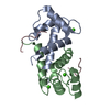
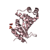
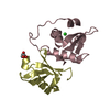
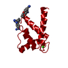
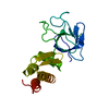
| Title | S100B-RSK1 crystal structure A' | ||||||
 Components Components |
| ||||||
 Keywords Keywords |  TRANSFERASE / Kinase / Signaling / Inhibitor / S100 TRANSFERASE / Kinase / Signaling / Inhibitor / S100 | ||||||
| Function / homology |  Function and homology informationregulation of translation in response to stress / CREB1 phosphorylation through NMDA receptor-mediated activation of RAS signaling / ribosomal protein S6 kinase activity / adaptive thermogenesis / CREB phosphorylation / hepatocyte proliferation / positive regulation of hepatic stellate cell activation / sympathetic neuron projection extension / RAGE receptor binding / Gastrin-CREB signalling pathway via PKC and MAPK ...regulation of translation in response to stress / CREB1 phosphorylation through NMDA receptor-mediated activation of RAS signaling / ribosomal protein S6 kinase activity / adaptive thermogenesis / CREB phosphorylation / hepatocyte proliferation / positive regulation of hepatic stellate cell activation / sympathetic neuron projection extension / RAGE receptor binding / Gastrin-CREB signalling pathway via PKC and MAPK / ion binding / RSK activation / S100 protein binding / negative regulation of TOR signaling / regulation of neuronal synaptic plasticity / ERK/MAPK targets / Recycling pathway of L1 / cysteine-type endopeptidase inhibitor activity involved in apoptotic process / TRAF6 mediated NF-kB activation / Advanced glycosylation endproduct receptor signaling / Nuclear signaling by ERBB4 / ruffle / positive regulation of neuron differentiation / protein serine/threonine/tyrosine kinase activity / axonogenesis / sarcoplasmic reticulum / central nervous system development / positive regulation of cell differentiation / TAK1-dependent IKK and NF-kappa-B activation / negative regulation of cysteine-type endopeptidase activity involved in apoptotic process / tau protein binding / memory / calcium-dependent protein binding / Senescence-Associated Secretory Phenotype (SASP) / positive regulation of cell growth / chemical synaptic transmission / positive regulation of canonical NF-kappaB signal transduction / learning or memory / non-specific serine/threonine protein kinase / cell adhesion / intracellular signal transduction / cell cycle / protein phosphorylation / intracellular membrane-bounded organelle / protein serine kinase activity / protein serine/threonine kinase activity / neuronal cell body / synapse / calcium ion binding / positive regulation of cell population proliferation / negative regulation of apoptotic process / perinuclear region of cytoplasm / positive regulation of DNA-templated transcription / magnesium ion binding / signal transduction / protein homodimerization activity / positive regulation of transcription by RNA polymerase II / extracellular space / zinc ion binding / extracellular region / nucleoplasm / ATP binding / identical protein binding / nucleus / cytosol / cytoplasm Function and homology informationregulation of translation in response to stress / CREB1 phosphorylation through NMDA receptor-mediated activation of RAS signaling / ribosomal protein S6 kinase activity / adaptive thermogenesis / CREB phosphorylation / hepatocyte proliferation / positive regulation of hepatic stellate cell activation / sympathetic neuron projection extension / RAGE receptor binding / Gastrin-CREB signalling pathway via PKC and MAPK ...regulation of translation in response to stress / CREB1 phosphorylation through NMDA receptor-mediated activation of RAS signaling / ribosomal protein S6 kinase activity / adaptive thermogenesis / CREB phosphorylation / hepatocyte proliferation / positive regulation of hepatic stellate cell activation / sympathetic neuron projection extension / RAGE receptor binding / Gastrin-CREB signalling pathway via PKC and MAPK / ion binding / RSK activation / S100 protein binding / negative regulation of TOR signaling / regulation of neuronal synaptic plasticity / ERK/MAPK targets / Recycling pathway of L1 / cysteine-type endopeptidase inhibitor activity involved in apoptotic process / TRAF6 mediated NF-kB activation / Advanced glycosylation endproduct receptor signaling / Nuclear signaling by ERBB4 / ruffle / positive regulation of neuron differentiation / protein serine/threonine/tyrosine kinase activity / axonogenesis / sarcoplasmic reticulum / central nervous system development / positive regulation of cell differentiation / TAK1-dependent IKK and NF-kappa-B activation / negative regulation of cysteine-type endopeptidase activity involved in apoptotic process / tau protein binding / memory / calcium-dependent protein binding / Senescence-Associated Secretory Phenotype (SASP) / positive regulation of cell growth / chemical synaptic transmission / positive regulation of canonical NF-kappaB signal transduction / learning or memory / non-specific serine/threonine protein kinase / cell adhesion / intracellular signal transduction / cell cycle / protein phosphorylation / intracellular membrane-bounded organelle / protein serine kinase activity / protein serine/threonine kinase activity / neuronal cell body / synapse / calcium ion binding / positive regulation of cell population proliferation / negative regulation of apoptotic process / perinuclear region of cytoplasm / positive regulation of DNA-templated transcription / magnesium ion binding / signal transduction / protein homodimerization activity / positive regulation of transcription by RNA polymerase II / extracellular space / zinc ion binding / extracellular region / nucleoplasm / ATP binding / identical protein binding / nucleus / cytosol / cytoplasmSimilarity search - Function | ||||||
| Biological species |  Homo sapiens (human) Homo sapiens (human) | ||||||
| Method | X-RAY DIFFRACTION / SYNCHROTRON / MOLECULAR REPLACEMENT / Resolution: 2.13 Å | ||||||
 Authors Authors | Gogl, G. / Nyitray, L. | ||||||
 Citation Citation | Journal: J.Biol.Chem. / Year: 2016 Title: Structural Basis of Ribosomal S6 Kinase 1 (RSK1) Inhibition by S100B Protein: MODULATION OF THE EXTRACELLULAR SIGNAL-REGULATED KINASE (ERK) SIGNALING CASCADE IN A CALCIUM-DEPENDENT WAY. Authors: Gogl, G. / Alexa, A. / Kiss, B. / Katona, G. / Kovacs, M. / Bodor, A. / Remenyi, A. / Nyitray, L. | ||||||
| History |
|
- Structure visualization
| Structure viewer | Molecule: MolmilJmol/JSmol |
|---|
- Downloads & links
-Download
| PDBx/mmCIF format | 5csi.cif.gz | 96.6 KB | Display | PDBx/mmCIF format |
|---|---|---|---|---|
| PDB format | pdb5csi.ent.gz | 73.1 KB | Display | PDB format |
| PDBx/mmJSON format | 5csi.json.gz | Tree view | PDBx/mmJSON format | |
| Others |  Other downloads Other downloads |
-Validation report
| Arichive directory | https://data.pdbj.org/pub/pdb/validation_reports/cs/5csiftp://data.pdbj.org/pub/pdb/validation_reports/cs/5csi | HTTPS FTP |
|---|
-Related structure data
| Related structure data |  5csfC 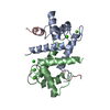 5csjC 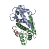 5csnC 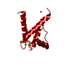 3cztS S: Starting model for refinement C: citing same article ( |
|---|---|
| Similar structure data |

-Links
 PDBj
PDBj


- Assembly
| Deposited unit | 
| ||||||||
|---|---|---|---|---|---|---|---|---|---|
| 1 |
| ||||||||
| Unit cell |
|
-Components
| #1: Protein | Mass: 11009.312 Da / Num. of mol.: 2 Source method: isolated from a genetically manipulated source Source: (gene. exp.) Homo sapiens (human) / Gene: S100B / Production host:  Escherichia coli (E. coli) / References: UniProt: P04271 Escherichia coli (E. coli) / References: UniProt: P04271#2: Protein/peptide | | Ribosome / S6K-alpha-1 / 90 kDa ribosomal protein S6 kinase 1 / p90S6K / MAP kinase-activated protein kinase ...S6K-alpha-1 / 90 kDa ribosomal protein S6 kinase 1 / p90S6K / MAP kinase-activated protein kinase 1a / MAPKAPK-1a / Ribosomal S6 kinase 1 / RSK-1Mass: 5260.097 Da / Num. of mol.: 1 / Fragment: UNP residues 689-735 Source method: isolated from a genetically manipulated source Source: (gene. exp.) Homo sapiens (human) / Gene: RPS6KA1, MAPKAPK1A, RSK1 / Production host: Escherichia coli (E. coli)References: UniProt: Q15418, non-specific serine/threonine protein kinase#3: Chemical | ChemComp-CA /   Mass: 40.078 Da / Num. of mol.: 4 / Source method: obtained synthetically / Formula: Ca Mass: 40.078 Da / Num. of mol.: 4 / Source method: obtained synthetically / Formula: Ca#4: Water | ChemComp-HOH / | Water Mass: 18.015 Da / Num. of mol.: 40 / Source method: isolated from a natural source / Formula: H2O Mass: 18.015 Da / Num. of mol.: 40 / Source method: isolated from a natural source / Formula: H2O |
|---|
-Experimental details
-Experiment
| Experiment | Method: X-RAY DIFFRACTION |
|---|
- Sample preparation
| Crystal | Density Matthews: 2.35 Å3/Da / Density % sol: 47.62 % |
|---|---|
| Crystal grow | Temperature: 296 K / Method: vapor diffusion, sitting drop / Details: 0.1 M Hepes 7, 150 mM NaCl, 20% PEG6000 |
-Data collection
| Diffraction | Mean temperature: 100 K |
|---|---|
| Diffraction source | Source: SYNCHROTRON / Site: ESRF  / Beamline: ID23-1 / Wavelength: 0.973 Å / Beamline: ID23-1 / Wavelength: 0.973 Å |
| Detector | Type: DECTRIS PILATUS 6M / Detector: PIXEL / Date: Nov 12, 2014 |
| Radiation | Protocol: SINGLE WAVELENGTH / Monochromatic (M) / Laue (L): M / Scattering type: x-ray |
| Radiation wavelength | Wavelength: 0.973 Å / Relative weight: 1 |
| Reflection | Resolution: 2.13→37.65 Å / Num. obs: 15160 / % possible obs: 99.9 % / Redundancy: 6.43 % / Rsym value: 0.055 / Net I/σ(I): 18.77 |
| Reflection shell | Resolution: 2.13→2.19 Å / Redundancy: 6.73 % / Mean I/σ(I) obs: 2.07 / Rsym value: 0.828 / % possible all: 100 |
- Processing
| Software |
| ||||||||||||||||||||||||||||||||||||||||||||||||||||||||||||||||||||||||||||||||||||||||||||||||||||||||||||||||||||||||||||||||||||||||||||||||||||||||||||||||||||||||||||||||||||||||||||||||||||||||||||||||||||||||||||||||||||||||||||||||||||||||||||||||||||||||||||||||||||||||||||||||||||||||||||||||||||||||||||||||||||||||||||||||||||||||||||||
|---|---|---|---|---|---|---|---|---|---|---|---|---|---|---|---|---|---|---|---|---|---|---|---|---|---|---|---|---|---|---|---|---|---|---|---|---|---|---|---|---|---|---|---|---|---|---|---|---|---|---|---|---|---|---|---|---|---|---|---|---|---|---|---|---|---|---|---|---|---|---|---|---|---|---|---|---|---|---|---|---|---|---|---|---|---|---|---|---|---|---|---|---|---|---|---|---|---|---|---|---|---|---|---|---|---|---|---|---|---|---|---|---|---|---|---|---|---|---|---|---|---|---|---|---|---|---|---|---|---|---|---|---|---|---|---|---|---|---|---|---|---|---|---|---|---|---|---|---|---|---|---|---|---|---|---|---|---|---|---|---|---|---|---|---|---|---|---|---|---|---|---|---|---|---|---|---|---|---|---|---|---|---|---|---|---|---|---|---|---|---|---|---|---|---|---|---|---|---|---|---|---|---|---|---|---|---|---|---|---|---|---|---|---|---|---|---|---|---|---|---|---|---|---|---|---|---|---|---|---|---|---|---|---|---|---|---|---|---|---|---|---|---|---|---|---|---|---|---|---|---|---|---|---|---|---|---|---|---|---|---|---|---|---|---|---|---|---|---|---|---|---|---|---|---|---|---|---|---|---|---|---|---|---|---|---|---|---|---|---|---|---|---|---|---|---|---|---|---|---|---|---|---|---|---|---|---|---|---|---|---|---|---|---|---|---|---|---|---|---|---|---|---|---|---|---|---|---|---|---|---|---|---|---|---|---|---|---|---|---|---|---|---|---|---|---|---|---|---|---|---|---|
| Refinement | Method to determine structure: MOLECULAR REPLACEMENT Starting model: 3CZT Resolution: 2.13→37.65 Å / SU ML: 0.28 / Cross valid method: FREE R-VALUE / σ(F): 1.36 / Phase error: 30.83 / Stereochemistry target values: ML
| ||||||||||||||||||||||||||||||||||||||||||||||||||||||||||||||||||||||||||||||||||||||||||||||||||||||||||||||||||||||||||||||||||||||||||||||||||||||||||||||||||||||||||||||||||||||||||||||||||||||||||||||||||||||||||||||||||||||||||||||||||||||||||||||||||||||||||||||||||||||||||||||||||||||||||||||||||||||||||||||||||||||||||||||||||||||||||||||
| Solvent computation | Shrinkage radii: 0.9 Å / VDW probe radii: 1.11 Å / Solvent model: FLAT BULK SOLVENT MODEL | ||||||||||||||||||||||||||||||||||||||||||||||||||||||||||||||||||||||||||||||||||||||||||||||||||||||||||||||||||||||||||||||||||||||||||||||||||||||||||||||||||||||||||||||||||||||||||||||||||||||||||||||||||||||||||||||||||||||||||||||||||||||||||||||||||||||||||||||||||||||||||||||||||||||||||||||||||||||||||||||||||||||||||||||||||||||||||||||
| Refinement step | Cycle: LAST / Resolution: 2.13→37.65 Å
| ||||||||||||||||||||||||||||||||||||||||||||||||||||||||||||||||||||||||||||||||||||||||||||||||||||||||||||||||||||||||||||||||||||||||||||||||||||||||||||||||||||||||||||||||||||||||||||||||||||||||||||||||||||||||||||||||||||||||||||||||||||||||||||||||||||||||||||||||||||||||||||||||||||||||||||||||||||||||||||||||||||||||||||||||||||||||||||||
| Refine LS restraints |
| ||||||||||||||||||||||||||||||||||||||||||||||||||||||||||||||||||||||||||||||||||||||||||||||||||||||||||||||||||||||||||||||||||||||||||||||||||||||||||||||||||||||||||||||||||||||||||||||||||||||||||||||||||||||||||||||||||||||||||||||||||||||||||||||||||||||||||||||||||||||||||||||||||||||||||||||||||||||||||||||||||||||||||||||||||||||||||||||
| LS refinement shell |
| ||||||||||||||||||||||||||||||||||||||||||||||||||||||||||||||||||||||||||||||||||||||||||||||||||||||||||||||||||||||||||||||||||||||||||||||||||||||||||||||||||||||||||||||||||||||||||||||||||||||||||||||||||||||||||||||||||||||||||||||||||||||||||||||||||||||||||||||||||||||||||||||||||||||||||||||||||||||||||||||||||||||||||||||||||||||||||||||
| Refinement TLS params. | Method: refined / Refine-ID: X-RAY DIFFRACTION
| ||||||||||||||||||||||||||||||||||||||||||||||||||||||||||||||||||||||||||||||||||||||||||||||||||||||||||||||||||||||||||||||||||||||||||||||||||||||||||||||||||||||||||||||||||||||||||||||||||||||||||||||||||||||||||||||||||||||||||||||||||||||||||||||||||||||||||||||||||||||||||||||||||||||||||||||||||||||||||||||||||||||||||||||||||||||||||||||
| Refinement TLS group |
|
