[English] 日本語
Yorodumi
- PDB-4e9m: NOD1 card domain with three disulfide-clinched, domain-swapped di... -

+
Open data


ID or keywords:

Loading...

-
Basic information

Entry
Database: PDB / ID: 4e9m
TitleNOD1 card domain with three disulfide-clinched, domain-swapped dimers in the asymmetric unit
ComponentsNucleotide-binding oligomerization domain-containing protein 1
KeywordsPROTEIN BINDING / INNATE IMMUNITY / RIPK2
Function / homology
Function and homology information


positive regulation of dendritic cell antigen processing and presentation / detection of biotic stimulus / positive regulation of xenophagy / xenophagy / nucleotide-binding oligomerization domain containing 1 signaling pathway / cellular response to muramyl dipeptide / CARD domain binding / peptidoglycan binding / positive regulation of macrophage cytokine production / pattern recognition receptor activity ...positive regulation of dendritic cell antigen processing and presentation / detection of biotic stimulus / positive regulation of xenophagy / xenophagy / nucleotide-binding oligomerization domain containing 1 signaling pathway / cellular response to muramyl dipeptide / CARD domain binding / peptidoglycan binding / positive regulation of macrophage cytokine production / pattern recognition receptor activity / pattern recognition receptor signaling pathway / cysteine-type endopeptidase activator activity involved in apoptotic process / positive regulation of cysteine-type endopeptidase activity involved in apoptotic process / phagocytic vesicle / JNK cascade / ERK1 and ERK2 cascade / detection of bacterium / response to endoplasmic reticulum stress / JNK (c-Jun kinases) phosphorylation and activation mediated by activated human TAK1 / ubiquitin binding / positive regulation of interleukin-1 beta production / activated TAK1 mediates p38 MAPK activation / positive regulation of interleukin-8 production / positive regulation of JNK cascade / TAK1-dependent IKK and NF-kappa-B activation / NOD1/2 Signaling Pathway / defense response / Interleukin-1 signaling / positive regulation of interleukin-6 production / positive regulation of non-canonical NF-kappaB signal transduction / activation of cysteine-type endopeptidase activity involved in apoptotic process / Ovarian tumor domain proteases / positive regulation of tumor necrosis factor production / positive regulation of NF-kappaB transcription factor activity / basolateral plasma membrane / positive regulation of canonical NF-kappaB signal transduction / defense response to Gram-negative bacterium / positive regulation of ERK1 and ERK2 cascade / intracellular signal transduction / defense response to Gram-positive bacterium / defense response to bacterium / inflammatory response / positive regulation of apoptotic process / apical plasma membrane / innate immune response / apoptotic process / protein-containing complex binding / SARS-CoV-2 activates/modulates innate and adaptive immune responses / signal transduction / protein homodimerization activity / ATP binding / identical protein binding / plasma membrane / cytosol / cytoplasm
Similarity search - Function
NACHT, LRR and PYD domains-containing protein, helical domain HD2 / NLRC4 helical domain HD2 / NOD2, winged helix domain / NOD2 winged helix domain / NACHT nucleoside triphosphatase / NACHT domain / NACHT-NTPase domain profile. / Death Domain, Fas / Death Domain, Fas / Leucine rich repeat, ribonuclease inhibitor type ...NACHT, LRR and PYD domains-containing protein, helical domain HD2 / NLRC4 helical domain HD2 / NOD2, winged helix domain / NOD2 winged helix domain / NACHT nucleoside triphosphatase / NACHT domain / NACHT-NTPase domain profile. / Death Domain, Fas / Death Domain, Fas / Leucine rich repeat, ribonuclease inhibitor type / Leucine Rich repeat / CARD domain / CARD caspase recruitment domain profile. / Caspase recruitment domain / Death-like domain superfamily / Leucine-rich repeat / Leucine-rich repeat domain superfamily / P-loop containing nucleoside triphosphate hydrolase / Orthogonal Bundle / Mainly Alpha
Similarity search - Domain/homology
CITRATE ANION / Nucleotide-binding oligomerization domain-containing protein 1
Similarity search - Component
Biological speciesHomo sapiens (human)
MethodX-RAY DIFFRACTION / SYNCHROTRON / MOLECULAR REPLACEMENT / Resolution: 2.15 Å
AuthorsBerry, E.A. / Huang, L.S. / Walker-Kopp, N.
CitationJournal: To be Published
Title: Crystal structure of nod1 card domain with three disulfide-clinched, domain-swapped dimers in the asymmetric unit
Authors: Berry, E.A. / Huang, L.S. / Walker-Kopp, N.
History
DepositionMar 21, 2012Deposition site: RCSB / Processing site: RCSB
Revision 1.0Apr 4, 2012Provider: repository / Type: Initial release
Revision 1.1Feb 6, 2013Group: Other
Revision 1.2Sep 13, 2023Group: Data collection / Database references ...Data collection / Database references / Derived calculations / Refinement description
Category: chem_comp_atom / chem_comp_bond ...chem_comp_atom / chem_comp_bond / database_2 / pdbx_initial_refinement_model / struct_ref_seq_dif / struct_site
Item: _database_2.pdbx_DOI / _database_2.pdbx_database_accession ..._database_2.pdbx_DOI / _database_2.pdbx_database_accession / _struct_ref_seq_dif.details / _struct_site.pdbx_auth_asym_id / _struct_site.pdbx_auth_comp_id / _struct_site.pdbx_auth_seq_id

-
Structure visualization

Structure viewerMolecule:
MolmilJmol/JSmol

Downloads & links

-
Assembly

Deposited unit
A: Nucleotide-binding oligomerization domain-containing protein 1
B: Nucleotide-binding oligomerization domain-containing protein 1
C: Nucleotide-binding oligomerization domain-containing protein 1
D: Nucleotide-binding oligomerization domain-containing protein 1
E: Nucleotide-binding oligomerization domain-containing protein 1
F: Nucleotide-binding oligomerization domain-containing protein 1
hetero molecules


Theoretical massNumber of molelcules
Total (without water)100,3227
Polymers100,1336
Non-polymers1891
Water1,58588
1
A: Nucleotide-binding oligomerization domain-containing protein 1


Theoretical massNumber of molelcules
Total (without water)16,6891
Polymers16,6891
Non-polymers00
Water181
TypeNameSymmetry operationNumber
identity operation1_555x,y,z1
2
B: Nucleotide-binding oligomerization domain-containing protein 1


Theoretical massNumber of molelcules
Total (without water)16,6891
Polymers16,6891
Non-polymers00
Water181
TypeNameSymmetry operationNumber
identity operation1_555x,y,z1
3
C: Nucleotide-binding oligomerization domain-containing protein 1


Theoretical massNumber of molelcules
Total (without water)16,6891
Polymers16,6891
Non-polymers00
Water181
TypeNameSymmetry operationNumber
identity operation1_555x,y,z1
4
D: Nucleotide-binding oligomerization domain-containing protein 1
hetero molecules


Theoretical massNumber of molelcules
Total (without water)16,8782
Polymers16,6891
Non-polymers1891
Water181
TypeNameSymmetry operationNumber
identity operation1_555x,y,z1
5
E: Nucleotide-binding oligomerization domain-containing protein 1


Theoretical massNumber of molelcules
Total (without water)16,6891
Polymers16,6891
Non-polymers00
Water181
TypeNameSymmetry operationNumber
identity operation1_555x,y,z1
6
F: Nucleotide-binding oligomerization domain-containing protein 1


Theoretical massNumber of molelcules
Total (without water)16,6891
Polymers16,6891
Non-polymers00
Water181
TypeNameSymmetry operationNumber
identity operation1_555x,y,z1
7
A: Nucleotide-binding oligomerization domain-containing protein 1
B: Nucleotide-binding oligomerization domain-containing protein 1


Theoretical massNumber of molelcules
Total (without water)33,3782
Polymers33,3782
Non-polymers00
Water362
TypeNameSymmetry operationNumber
identity operation1_555x,y,z1
Buried area3190 Å2
ΔGint-22 kcal/mol
Surface area9810 Å2
MethodPISA
8
C: Nucleotide-binding oligomerization domain-containing protein 1
D: Nucleotide-binding oligomerization domain-containing protein 1
hetero molecules


Theoretical massNumber of molelcules
Total (without water)33,5673
Polymers33,3782
Non-polymers1891
Water362
TypeNameSymmetry operationNumber
identity operation1_555x,y,z1
Buried area3010 Å2
ΔGint-17 kcal/mol
Surface area9470 Å2
MethodPISA
9
E: Nucleotide-binding oligomerization domain-containing protein 1
F: Nucleotide-binding oligomerization domain-containing protein 1


Theoretical massNumber of molelcules
Total (without water)33,3782
Polymers33,3782
Non-polymers00
Water362
TypeNameSymmetry operationNumber
identity operation1_555x,y,z1
Buried area2860 Å2
ΔGint-19 kcal/mol
Surface area9520 Å2
MethodPISA
Unit cell
Length a, b, c (Å)82.846, 85.335, 113.682
Angle α, β, γ (deg.)90.00, 90.00, 90.00
Int Tables number19
Space group name H-MP212121
Details
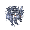
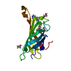
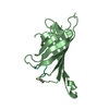
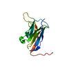
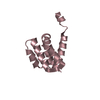
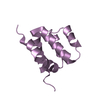
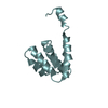
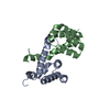
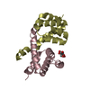
IT IS NOT KNOWN WHETHER THE DOMAIN-SWAPPED DIMERS SEEN HERE HAVE PHYSIOLOGICAL FUNCTION. CARD DOMAINS ARE KNOWN TO FORM HETERODIMERS WITH OTHER CARDS, BUT THIS IS NOT BELIEVED TO BE BY A DOMAIN-SWAPPING MECHANISM

-
Components

#1: Protein
Nucleotide-binding oligomerization domain-containing protein 1 / Caspase recruitment domain-containing protein 4


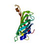
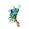
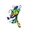
Mass: 16688.891 Da / Num. of mol.: 6 / Fragment: CARD DOMAIN
Source method: isolated from a genetically manipulated source
Source: (gene. exp.) Homo sapiens (human) / Gene: CARD4, NOD1 / Production host: Escherichia coli (E. coli) / Strain (production host): BL21 / References: UniProt: Q9Y239
#2: Chemical ChemComp-FLC / CITRATE ANION / Citric acid


Mass: 189.100 Da / Num. of mol.: 1 / Source method: obtained synthetically / Formula: C6H5O7
#3: Water ChemComp-HOH / water / Water


Mass: 18.015 Da / Num. of mol.: 88 / Source method: isolated from a natural source / Formula: H2O

-
Experimental details

-
Experiment

ExperimentMethod: X-RAY DIFFRACTION / Number of used crystals: 1

-
Sample preparation

CrystalDensity Matthews: 2.01 Å3/Da / Density % sol: 38.7 %
Crystal growTemperature: 288 K / Method: vapor diffusion, hanging drop / pH: 5.2
Details: 2.07 M AMMONIUM CITRATE (DIBASIC), 6.7% (W/V) PEG3350, 10 mM TRIS-HCL FINAL PH ~5.2, VAPOR DIFFUSION, HANGING DROP, temperature 288K

-
Data collection

DiffractionMean temperature: 100 K
Diffraction sourceSource: SYNCHROTRON / Site: CHESS / Beamline: A1 / Wavelength: 0.977 Å
DetectorType: ADSC QUANTUM 210 / Detector: CCD / Date: May 1, 2011
RadiationMonochromator: double, Horizontal focusing 5.05 asymmetric cut Si(111)
Protocol: SINGLE WAVELENGTH / Monochromatic (M) / Laue (L): M / Scattering type: x-ray
Radiation wavelengthWavelength: 0.977 Å / Relative weight: 1
ReflectionResolution: 2.15→25 Å / Num. all: 34341 / Num. obs: 34341 / % possible obs: 75.9 % / Observed criterion σ(I): -3 / Redundancy: 3.6 % / Biso Wilson estimate: 39.4 Å2 / Rsym value: 0.084 / Net I/σ(I): 9.66
Reflection shell
Resolution (Å)Redundancy (%)Mean I/σ(I) obsRsym valueDiffraction-ID% possible all
2.15-2.191.30.790.54517
2.19-2.231.41.120.41114.3
2.55-2.622.93.040.38191.7
4.05-4.634.217.60.056199.2
5.83-503.9170.039197.8

-
Processing

Software
NameVersionClassification
ADSCQuantumdata collection
PHASERphasing
CNS1.1refinement
DENZOdata reduction
SCALEPACKdata scaling
RefinementMethod to determine structure: MOLECULAR REPLACEMENT
Starting model: PDB ENTRY 2NZ7
Resolution: 2.15→24.84 Å / Rfactor Rfree error: 0.007 / Data cutoff high absF: 2255943.04 / Data cutoff low absF: 0 / Isotropic thermal model: RESTRAINED / Cross valid method: THROUGHOUT / σ(F): 0 / Stereochemistry target values: Engh & Huber
Details: UP TO 6-FOLD NCS RESTRAINTS ON COORDINATES WERE USED IN 9 NCS GROUPS, WITH NUMEROUS EXCEPTIONS FOR RESIDUES WITH DIFFERENT CONFORMATIONS. NO NCS RESTRAINTS WERE IMPOSED ON BFACTORS. Low ...Details: UP TO 6-FOLD NCS RESTRAINTS ON COORDINATES WERE USED IN 9 NCS GROUPS, WITH NUMEROUS EXCEPTIONS FOR RESIDUES WITH DIFFERENT CONFORMATIONS. NO NCS RESTRAINTS WERE IMPOSED ON BFACTORS. Low completeness is due to use of data in corners of detector. sfcheck plot of completenes vs resolution shows good completeness to 2.6 Angstrom.
RfactorNum. reflection% reflectionSelection details
Rfree0.275 1701 5 %RANDOM
Rwork0.245 ---
obs0.245 34213 76.8 %-
all-34213 --
Solvent computationSolvent model: FLAT MODEL / Bsol: 49.3675 Å2 / ksol: 0.361962 e/Å3
Displacement parametersBiso mean: 51.7 Å2
Baniso -1Baniso -2Baniso -3
1-1.52 Å20 Å20 Å2
2---2.56 Å20 Å2
3---1.03 Å2
Refine analyze
FreeObs
Luzzati coordinate error0.39 Å0.33 Å
Luzzati d res low-5 Å
Luzzati sigma a0.33 Å0.3 Å
Refinement stepCycle: LAST / Resolution: 2.15→24.84 Å
ProteinNucleic acidLigandSolventTotal
Num. atoms4540 0 13 88 4641
Refine LS restraints
Refine-IDTypeDev idealDev ideal target
X-RAY DIFFRACTIONc_bond_d0.006
X-RAY DIFFRACTIONc_bond_d_na
X-RAY DIFFRACTIONc_bond_d_prot
X-RAY DIFFRACTIONc_angle_d
X-RAY DIFFRACTIONc_angle_d_na
X-RAY DIFFRACTIONc_angle_d_prot
X-RAY DIFFRACTIONc_angle_deg1.2
X-RAY DIFFRACTIONc_angle_deg_na
X-RAY DIFFRACTIONc_angle_deg_prot
X-RAY DIFFRACTIONc_dihedral_angle_d18.9
X-RAY DIFFRACTIONc_dihedral_angle_d_na
X-RAY DIFFRACTIONc_dihedral_angle_d_prot
X-RAY DIFFRACTIONc_improper_angle_d0.84
X-RAY DIFFRACTIONc_improper_angle_d_na
X-RAY DIFFRACTIONc_improper_angle_d_prot
X-RAY DIFFRACTIONc_mcbond_it1.421.5
X-RAY DIFFRACTIONc_mcangle_it2.372
X-RAY DIFFRACTIONc_scbond_it2.252
X-RAY DIFFRACTIONc_scangle_it3.42.5
Refine LS restraints NCSNCS model details: RESTRAINED
LS refinement shellResolution: 2.15→2.26 Å / Rfactor Rfree error: 0.055 / Total num. of bins used: 7
RfactorNum. reflection% reflection
Rfree0.406 54 5.2 %
Rwork0.352 987 -
obs--16.6 %
Xplor file
Refine-IDSerial noParam file
X-RAY DIFFRACTION1protein_rep.param
X-RAY DIFFRACTION2flc.par
X-RAY DIFFRACTION4water.param

+
About Yorodumi

-
News

-
Feb 9, 2022. New format data for meta-information of EMDB entries

New format data for meta-information of EMDB entries

  • Version 3 of the EMDB header file is now the official format.
  • The previous official version 1.9 will be removed from the archive.

Related info.:EMDB header

External links:wwPDB to switch to version 3 of the EMDB data model

-
Aug 12, 2020. Covid-19 info

Covid-19 info

URL: https://pdbj.org/emnavi/covid19.php

New page: Covid-19 featured information page in EM Navigator.

Related info.:Covid-19 info / Mar 5, 2020. Novel coronavirus structure data

+
Mar 5, 2020. Novel coronavirus structure data

Novel coronavirus structure data

Related info.:Yorodumi Speices / Aug 12, 2020. Covid-19 info

External links:COVID-19 featured content - PDBj / Molecule of the Month (242):Coronavirus Proteases

+
Jan 31, 2019. EMDB accession codes are about to change! (news from PDBe EMDB page)

EMDB accession codes are about to change! (news from PDBe EMDB page)

  • The allocation of 4 digits for EMDB accession codes will soon come to an end. Whilst these codes will remain in use, new EMDB accession codes will include an additional digit and will expand incrementally as the available range of codes is exhausted. The current 4-digit format prefixed with “EMD-” (i.e. EMD-XXXX) will advance to a 5-digit format (i.e. EMD-XXXXX), and so on. It is currently estimated that the 4-digit codes will be depleted around Spring 2019, at which point the 5-digit format will come into force.
  • The EM Navigator/Yorodumi systems omit the EMD- prefix.

Related info.:Q: What is EMD? / ID/Accession-code notation in Yorodumi/EM Navigator

External links:EMDB Accession Codes are Changing Soon! / Contact to PDBj

+
Jul 12, 2017. Major update of PDB

Major update of PDB

  • wwPDB released updated PDB data conforming to the new PDBx/mmCIF dictionary.
  • This is a major update changing the version number from 4 to 5, and with Remediation, in which all the entries are updated.
  • In this update, many items about electron microscopy experimental information are reorganized (e.g. em_software).
  • Now, EM Navigator and Yorodumi are based on the updated data.

External links:wwPDB Remediation / Enriched Model Files Conforming to OneDep Data Standards Now Available in the PDB FTP Archive

-
Yorodumi

Thousand views of thousand structures

  • Yorodumi is a browser for structure data from EMDB, PDB, SASBDB, etc.
  • This page is also the successor to EM Navigator detail page, and also detail information page/front-end page for Omokage search.
  • The word "yorodu" (or yorozu) is an old Japanese word meaning "ten thousand". "mi" (miru) is to see.

Related info.:EMDB / PDB / SASBDB / Comparison of 3 databanks / Yorodumi Search / Aug 31, 2016. New EM Navigator & Yorodumi / Yorodumi Papers / Jmol/JSmol / Function and homology information / Changes in new EM Navigator and Yorodumi

Read more