 Movie
Movie Controller


+ Open data

- Basic information
| Entry | Database: PDB / ID: 3re6 | ||||||
|---|---|---|---|---|---|---|---|
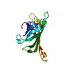
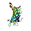
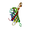
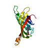
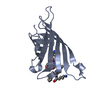
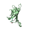
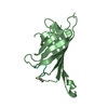
| Title | Crystal structure of R4-6 streptavidin | ||||||
 Components Components | Streptavidin | ||||||
 Keywords Keywords | BIOTIN BINDING PROTEIN / Streptavidin variants / improved desthiobiotin binding / opened loop destabilization | ||||||
| Function / homology |  Function and homology information Function and homology information | ||||||
| Biological species |  Streptomyces avidinii (bacteria) Streptomyces avidinii (bacteria) | ||||||
| Method | X-RAY DIFFRACTION / SYNCHROTRON / MOLECULAR REPLACEMENT / molecular replacement / Resolution: 1.823 Å | ||||||
 Authors Authors | Malashkevich, V.N. / Magalhaes, M. / Czecster, C.M. / Guan, R. / Levy, M. / Almo, S.C. | ||||||
 Citation Citation | Journal: Protein Sci. / Year: 2011 Title: Evolved streptavidin mutants reveal key role of loop residue in high-affinity binding. Authors: Magalhaes, M.L. / Czekster, C.M. / Guan, R. / Malashkevich, V.N. / Almo, S.C. / Levy, M. | ||||||
| History |
|
- Structure visualization
| Structure viewer | Molecule: MolmilJmol/JSmol |
|---|
- Downloads & links
-Download
| PDBx/mmCIF format | 3re6.cif.gz | 61.6 KB | Display | PDBx/mmCIF format |
|---|---|---|---|---|
| PDB format | pdb3re6.ent.gz | 44.7 KB | Display | PDB format |
| PDBx/mmJSON format | 3re6.json.gz | Tree view | PDBx/mmJSON format | |
| Others |  Other downloads Other downloads |
-Validation report
| Arichive directory | https://data.pdbj.org/pub/pdb/validation_reports/re/3re6ftp://data.pdbj.org/pub/pdb/validation_reports/re/3re6 | HTTPS FTP |
|---|
-Related structure data
| Related structure data |  3rdmC 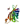 3rdoC  3rdqC 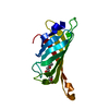 3rdsSC  3rduC  3rdxC 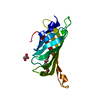 3re5C C: citing same article ( S: Starting model for refinement |
|---|---|
| Similar structure data |


-Links
 PDBj
PDBj- Assembly
| Deposited unit | 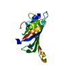
| |||||||||
|---|---|---|---|---|---|---|---|---|---|---|
| 1 |
| |||||||||
| 2 | 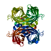
| |||||||||
| Unit cell |
| |||||||||
| Components on special symmetry positions |
|
-Components
| #1: Protein | Mass: 15996.406 Da / Num. of mol.: 1 / Fragment: UNP Residues 37-164 / Mutation: T90S,W108V,L110T,F29L,R53S Source method: isolated from a genetically manipulated source Source: (gene. exp.) Streptomyces avidinii (bacteria) / Plasmid: pCR2.1-TOPO / Production host: Escherichia coli (E. coli) / Strain (production host): BL21(DE3)CODON+RIL / References: UniProt: P22629 |
|---|---|
| #2: Chemical | ChemComp-GOL / Glycerol  Mass: 92.094 Da / Num. of mol.: 1 / Source method: obtained synthetically / Formula: C3H8O3 Mass: 92.094 Da / Num. of mol.: 1 / Source method: obtained synthetically / Formula: C3H8O3 |
| #3: Water | ChemComp-HOH / Water Mass: 18.015 Da / Num. of mol.: 97 / Source method: isolated from a natural source / Formula: H2O Mass: 18.015 Da / Num. of mol.: 97 / Source method: isolated from a natural source / Formula: H2O |
-Experimental details
-Experiment
| Experiment | Method: X-RAY DIFFRACTION / Number of used crystals: 1 |
|---|
- Sample preparation
| Crystal | Density Matthews: 2.24 Å3/Da / Density % sol: 45.06 % |
|---|---|
| Crystal grow | Temperature: 298 K / Method: vapor diffusion, sitting drop / pH: 6.5 Details: 18% PEG 8000, 0.1 M Na-cacodylate, pH 6.5, 0.2 M Ca-acetate, VAPOR DIFFUSION, SITTING DROP, temperature 298K |
-Data collection
| Diffraction | Mean temperature: 100 K |
|---|---|
| Diffraction source | Source: SYNCHROTRON / Site: NSLS  / Beamline: X29A / Wavelength: 0.9791 Å / Beamline: X29A / Wavelength: 0.9791 Å |
| Detector | Type: ADSC QUANTUM 315 / Detector: CCD / Date: Jun 30, 2010 |
| Radiation | Protocol: SINGLE WAVELENGTH / Monochromatic (M) / Laue (L): M / Scattering type: x-ray |
| Radiation wavelength | Wavelength: 0.9791 Å / Relative weight: 1 |
| Reflection | Resolution: 1.82→20 Å / Num. obs: 13522 / % possible obs: 99.6 % / Redundancy: 13.5 % / Rmerge(I) obs: 0.093 |
| Reflection shell | Resolution: 1.82→1.85 Å / Redundancy: 13 % / Rmerge(I) obs: 0.539 / Num. unique all: 617 / Rsym value: 0.539 / % possible all: 92 |
-Phasing
| Phasing | Method: molecular replacement |
|---|
- Processing
| Software |
| |||||||||||||||||||||||||||||||||||||||||||||||||||||||||||||||||
|---|---|---|---|---|---|---|---|---|---|---|---|---|---|---|---|---|---|---|---|---|---|---|---|---|---|---|---|---|---|---|---|---|---|---|---|---|---|---|---|---|---|---|---|---|---|---|---|---|---|---|---|---|---|---|---|---|---|---|---|---|---|---|---|---|---|---|
| Refinement | Method to determine structure: MOLECULAR REPLACEMENT Starting model: PDB ENTRY 3RDS Resolution: 1.823→19.79 Å / Cor.coef. Fo:Fc: 0.953 / Cor.coef. Fo:Fc free: 0.92 / WRfactor Rfree: 0.22 / WRfactor Rwork: 0.1812 / Occupancy max: 1 / Occupancy min: 0.3 / FOM work R set: 0.8721 / SU B: 4.415 / SU ML: 0.064 / SU R Cruickshank DPI: 0.1194 / SU Rfree: 0.1214 / Cross valid method: THROUGHOUT / σ(F): 0 / ESU R: 0.119 / ESU R Free: 0.121 / Stereochemistry target values: MAXIMUM LIKELIHOOD Details: HYDROGENS HAVE BEEN ADDED IN THE RIDING POSITIONS U VALUES : RESIDUAL ONLY
| |||||||||||||||||||||||||||||||||||||||||||||||||||||||||||||||||
| Solvent computation | Ion probe radii: 0.8 Å / Shrinkage radii: 0.8 Å / VDW probe radii: 1.4 Å / Solvent model: BABINET MODEL WITH MASK | |||||||||||||||||||||||||||||||||||||||||||||||||||||||||||||||||
| Displacement parameters | Biso max: 65.17 Å2 / Biso mean: 25.94 Å2 / Biso min: 11.92 Å2
| |||||||||||||||||||||||||||||||||||||||||||||||||||||||||||||||||
| Refinement step | Cycle: LAST / Resolution: 1.823→19.79 Å
| |||||||||||||||||||||||||||||||||||||||||||||||||||||||||||||||||
| Refine LS restraints |
| |||||||||||||||||||||||||||||||||||||||||||||||||||||||||||||||||
| LS refinement shell | Resolution: 1.823→1.87 Å / Total num. of bins used: 20
| |||||||||||||||||||||||||||||||||||||||||||||||||||||||||||||||||
| Refinement TLS params. | Method: refined / Origin x: -13.4696 Å / Origin y: -24.5671 Å / Origin z: -2.1331 Å
|
