 Movie
Movie Controller


+ Open data

- Basic information
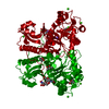
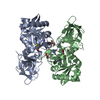
| Entry | Database: PDB / ID: 5cre | ||||||
|---|---|---|---|---|---|---|---|
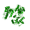
| Title | Human skeletal calsequestrin, D210G mutant low-calcium complex | ||||||
 Components Components | Calsequestrin-1 | ||||||
 Keywords Keywords | Calcium binding protein / Calsequestrin Calcium-binding protein | ||||||
| Function / homology |  Function and homology information Function and homology informationterminal cisterna lumen / positive regulation of store-operated calcium channel activity / regulation of skeletal muscle contraction by regulation of release of sequestered calcium ion / regulation of store-operated calcium entry / response to denervation involved in regulation of muscle adaptation / sarcoplasmic reticulum lumen / endoplasmic reticulum organization / sarcomere organization / smooth endoplasmic reticulum / protein polymerization ...terminal cisterna lumen / positive regulation of store-operated calcium channel activity / regulation of skeletal muscle contraction by regulation of release of sequestered calcium ion / regulation of store-operated calcium entry / response to denervation involved in regulation of muscle adaptation / sarcoplasmic reticulum lumen / endoplasmic reticulum organization / sarcomere organization / smooth endoplasmic reticulum / protein polymerization / skeletal muscle tissue development / Ion homeostasis / sarcoplasmic reticulum membrane / T-tubule / positive regulation of release of sequestered calcium ion into cytosol / sarcoplasmic reticulum / response to organic substance / Stimuli-sensing channels / Z disc / response to heat / mitochondrial matrix / calcium ion binding / endoplasmic reticulum / mitochondrion / identical protein bindingSimilarity search - Function | ||||||
| Biological species |  Homo sapiens (human) Homo sapiens (human) | ||||||
| Method | X-RAY DIFFRACTION / SYNCHROTRON / MOLECULAR REPLACEMENT / Resolution: 3.315 Å | ||||||
 Authors Authors | Lewis, K.M. / Ronish, L.A. / Kang, C. | ||||||
 Citation Citation | Journal: J.Biol.Chem. / Year: 2015 Title: Characterization of Two Human Skeletal Calsequestrin Mutants Implicated in Malignant Hyperthermia and Vacuolar Aggregate Myopathy. Authors: Lewis, K.M. / Ronish, L.A. / Rios, E. / Kang, C. | ||||||
| History |
|
- Structure visualization
| Structure viewer | Molecule: MolmilJmol/JSmol |
|---|
- Downloads & links
-Download
| PDBx/mmCIF format | 5cre.cif.gz | 83 KB | Display | PDBx/mmCIF format |
|---|---|---|---|---|
| PDB format | pdb5cre.ent.gz | 59.9 KB | Display | PDB format |
| PDBx/mmJSON format | 5cre.json.gz | Tree view | PDBx/mmJSON format | |
| Others |  Other downloads Other downloads |
-Validation report
| Arichive directory | https://data.pdbj.org/pub/pdb/validation_reports/cr/5creftp://data.pdbj.org/pub/pdb/validation_reports/cr/5cre | HTTPS FTP |
|---|
-Related structure data
| Related structure data | 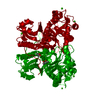 5crdSC  5crgC  5crhC C: citing same article ( S: Starting model for refinement |
|---|---|
| Similar structure data |


-Links
 PDBj
PDBj


- Assembly
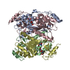
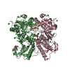
| Deposited unit | 
| ||||||||
|---|---|---|---|---|---|---|---|---|---|
| 1 | 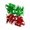
| ||||||||
| Unit cell |
|
-Components
| #1: Protein | / Calmitine / Calsequestrin / skeletal muscle isoform Mass: 41664.543 Da / Num. of mol.: 1 / Fragment: residues 35-396 / Mutation: D210G Source method: isolated from a genetically manipulated source Source: (gene. exp.) Homo sapiens (human) / Gene: CASQ1, CASQ / Production host:  Escherichia coli (E. coli) / References: UniProt: P31415 Escherichia coli (E. coli) / References: UniProt: P31415 | ||
|---|---|---|---|
| #2: Chemical |   Mass: 40.078 Da / Num. of mol.: 2 / Source method: obtained synthetically / Formula: Ca Mass: 40.078 Da / Num. of mol.: 2 / Source method: obtained synthetically / Formula: Ca#3: Chemical | 2-Methyl-2,4-pentanediol  Mass: 118.174 Da / Num. of mol.: 2 / Source method: obtained synthetically / Formula: C6H14O2 / Comment: precipitant*YM Mass: 118.174 Da / Num. of mol.: 2 / Source method: obtained synthetically / Formula: C6H14O2 / Comment: precipitant*YM |
-Experimental details
-Experiment
| Experiment | Method: X-RAY DIFFRACTION |
|---|
- Sample preparation
| Crystal | Density Matthews: 3.05 Å3/Da / Density % sol: 59.72 % |
|---|---|
| Crystal grow | Temperature: 277 K / Method: vapor diffusion, hanging drop Details: 0.1 M MOPS, 27.5 % (v/v) 2-methyl-2,4-pentanediol, 0.2 M NaCl |
-Data collection
| Diffraction | Mean temperature: 80 K |
|---|---|
| Diffraction source | Source: SYNCHROTRON / Site: ALS  / Beamline: 8.2.1 / Wavelength: 1 Å / Beamline: 8.2.1 / Wavelength: 1 Å |
| Detector | Type: ADSC QUANTUM 315r / Detector: CCD / Date: Feb 12, 2015 |
| Radiation | Protocol: SINGLE WAVELENGTH / Monochromatic (M) / Laue (L): M / Scattering type: x-ray |
| Radiation wavelength | Wavelength: 1 Å / Relative weight: 1 |
| Reflection | Resolution: 3.315→44.716 Å / Num. obs: 7649 / % possible obs: 99.62 % / Redundancy: 7 % / Net I/σ(I): 31.4 |
- Processing
| Software |
| ||||||||||||||||||||||||||||||||||||||||||
|---|---|---|---|---|---|---|---|---|---|---|---|---|---|---|---|---|---|---|---|---|---|---|---|---|---|---|---|---|---|---|---|---|---|---|---|---|---|---|---|---|---|---|---|
| Refinement | Method to determine structure: MOLECULAR REPLACEMENT Starting model: 5CRD Resolution: 3.315→44.716 Å / SU ML: 0.37 / Cross valid method: FREE R-VALUE / σ(F): 1.36 / Phase error: 29.61 / Stereochemistry target values: ML
| ||||||||||||||||||||||||||||||||||||||||||
| Solvent computation | Shrinkage radii: 0.9 Å / VDW probe radii: 1.11 Å / Solvent model: FLAT BULK SOLVENT MODEL | ||||||||||||||||||||||||||||||||||||||||||
| Refinement step | Cycle: LAST / Resolution: 3.315→44.716 Å
| ||||||||||||||||||||||||||||||||||||||||||
| Refine LS restraints |
| ||||||||||||||||||||||||||||||||||||||||||
| LS refinement shell |
|
