 Movie
Movie Controller


+ Open data

- Basic information
| Entry | Database: PDB / ID: 2vaf | |||||||||
|---|---|---|---|---|---|---|---|---|---|---|
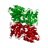
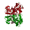
| Title | Crystal structure of Human Cardiac Calsequestrin | |||||||||
 Components Components | CALSEQUESTRIN-2 | |||||||||
 Keywords Keywords | METAL BINDING PROTEIN / CALCIUM / GLYCOPROTEIN / POLYMORPHISM / MUSCLE PROTEIN / DISEASE MUTATION / SARCOPLASMIC RETICULUM / CRYSTAL STRUCTURE HUMAN CARDIAC CALSEQUESTRIN / METAL-BINDING PROTEIN | |||||||||
| Function / homology |  Function and homology information Function and homology informationcalcium ion sequestering activity / negative regulation of potassium ion transmembrane transporter activity / regulation of cell communication by electrical coupling / sequestering of calcium ion / junctional sarcoplasmic reticulum membrane / Purkinje myocyte to ventricular cardiac muscle cell signaling / sarcoplasmic reticulum lumen / regulation of membrane repolarization / ion binding / cellular response to caffeine ...calcium ion sequestering activity / negative regulation of potassium ion transmembrane transporter activity / regulation of cell communication by electrical coupling / sequestering of calcium ion / junctional sarcoplasmic reticulum membrane / Purkinje myocyte to ventricular cardiac muscle cell signaling / sarcoplasmic reticulum lumen / regulation of membrane repolarization / ion binding / cellular response to caffeine / negative regulation of potassium ion transport / protein polymerization / detection of calcium ion / striated muscle contraction / negative regulation of ryanodine-sensitive calcium-release channel activity / regulation of cardiac muscle contraction by regulation of the release of sequestered calcium ion / regulation of release of sequestered calcium ion into cytosol by sarcoplasmic reticulum / cardiac muscle contraction / Ion homeostasis / sarcoplasmic reticulum membrane / calcium channel complex / regulation of heart rate / sarcoplasmic reticulum / intracellular calcium ion homeostasis / Stimuli-sensing channels / Z disc / calcium-dependent protein binding / calcium ion binding / protein homodimerization activity / cytoplasmSimilarity search - Function | |||||||||
| Biological species |  HOMO SAPIENS (human) HOMO SAPIENS (human) | |||||||||
| Method | X-RAY DIFFRACTION / SYNCHROTRON / MOLECULAR REPLACEMENT / Resolution: 3.8 Å | |||||||||
 Authors Authors | Kim, E. / Youn, B. / Kemper, L. / Campbell, C. / Milting, H. / Varsanyi, M. / Kang, C. | |||||||||
 Citation Citation | Journal: J.Mol.Biol. / Year: 2007 Title: Characterization of Human Cardiac Calsequestrin and its Deleterious Mutants. Authors: Kim, E. / Youn, B. / Kemper, L. / Campbell, C. / Milting, H. / Varsanyi, M. / Kang, C. | |||||||||
| History |
|
- Structure visualization
| Structure viewer | Molecule: MolmilJmol/JSmol |
|---|
- Downloads & links
-Download
| PDBx/mmCIF format | 2vaf.cif.gz | 84.2 KB | Display | PDBx/mmCIF format |
|---|---|---|---|---|
| PDB format | pdb2vaf.ent.gz | 63.4 KB | Display | PDB format |
| PDBx/mmJSON format | 2vaf.json.gz | Tree view | PDBx/mmJSON format | |
| Others |  Other downloads Other downloads |
-Validation report
| Arichive directory | https://data.pdbj.org/pub/pdb/validation_reports/va/2vafftp://data.pdbj.org/pub/pdb/validation_reports/va/2vaf | HTTPS FTP |
|---|
-Related structure data
| Related structure data |  1sjiS S: Starting model for refinement |
|---|---|
| Similar structure data |


-Links
 PDBj
PDBj


- Assembly
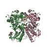
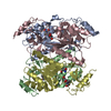
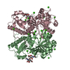
| Deposited unit | 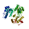
| ||||||||
|---|---|---|---|---|---|---|---|---|---|
| 1 | 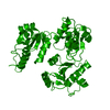
| ||||||||
| Unit cell |
|
-Components
| #1: Protein | / CALSEQUESTRIN / CARDIAC MUSCLE ISOFORM / HUMAN CARDIAC CALSEQUESTRIN Mass: 43997.586 Da / Num. of mol.: 1 / Fragment: RESIDUES 22-399 Source method: isolated from a genetically manipulated source Source: (gene. exp.) HOMO SAPIENS (human) / Organ: HEART / Production host:  ESCHERICHIA COLI (E. coli) / Strain (production host): BL21(DE3) / References: UniProt: O14958 ESCHERICHIA COLI (E. coli) / Strain (production host): BL21(DE3) / References: UniProt: O14958 |
|---|
-Experimental details
-Experiment
| Experiment | Method: X-RAY DIFFRACTION |
|---|
- Sample preparation
| Crystal | Density Matthews: 8.02 Å3/Da / Density % sol: 84.55 % / Description: NONE |
|---|
-Data collection
| Diffraction | Mean temperature: 277 K |
|---|---|
| Diffraction source | Source: SYNCHROTRON / Site: ALS  / Beamline: 8.2.1 / Wavelength: 1.07812 / Beamline: 8.2.1 / Wavelength: 1.07812 |
| Detector | Type: ADSC CCD / Detector: CCD |
| Radiation | Protocol: SINGLE WAVELENGTH / Monochromatic (M) / Laue (L): M / Scattering type: x-ray |
| Radiation wavelength | Wavelength: 1.07812 Å / Relative weight: 1 |
| Reflection | Resolution: 3.8→50 Å / Num. obs: 11522 / % possible obs: 88.7 % / Observed criterion σ(I): 2 / Redundancy: 8.4 % / Rmerge(I) obs: 0.09 / Net I/σ(I): 4.56 |
- Processing
| Software |
| ||||||||||||||||||||||||||||||||||||||||||||||||||||||||||||
|---|---|---|---|---|---|---|---|---|---|---|---|---|---|---|---|---|---|---|---|---|---|---|---|---|---|---|---|---|---|---|---|---|---|---|---|---|---|---|---|---|---|---|---|---|---|---|---|---|---|---|---|---|---|---|---|---|---|---|---|---|---|
| Refinement | Method to determine structure: MOLECULAR REPLACEMENT Starting model: PDB ENTRY 1SJI Resolution: 3.8→15 Å / Cross valid method: THROUGHOUT / σ(F): 2
| ||||||||||||||||||||||||||||||||||||||||||||||||||||||||||||
| Refinement step | Cycle: LAST / Resolution: 3.8→15 Å
| ||||||||||||||||||||||||||||||||||||||||||||||||||||||||||||
| Refine LS restraints |
| ||||||||||||||||||||||||||||||||||||||||||||||||||||||||||||
| Xplor file | Serial no: 1 / Param file: PARAM19X.PRO / Topol file: TOPH19X.PRO |
