 Movie
Movie Controller


+ Open data

- Basic information
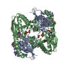
| Entry | Database: PDB / ID: 3tfi | ||||||
|---|---|---|---|---|---|---|---|
| Title | DMSP-dependent demethylase from P. ubique - with substrate DMSP | ||||||
 Components Components | GcvT-like Aminomethyltransferase protein | ||||||
 Keywords Keywords |  TRANSFERASE / demethylase / THF TRANSFERASE / demethylase / THF | ||||||
| Function / homology |  Function and homology information Function and homology informationdimethylsulfoniopropionate demethylase / methyltransferase activity / methylationSimilarity search - Function | ||||||
| Biological species |  Candidatus Pelagibacter ubique (bacteria) Candidatus Pelagibacter ubique (bacteria) | ||||||
| Method | X-RAY DIFFRACTION / SYNCHROTRON / MOLECULAR REPLACEMENT / Resolution: 1.6 Å | ||||||
 Authors Authors | Schuller, D.J. / Reisch, C.R. / Moran, M.A. / Whitman, W.B. / Lanzilotta, W.N. | ||||||
 Citation Citation | Journal: Protein Sci. / Year: 2012 Title: Structures of dimethylsulfoniopropionate-dependent demethylase from the marine organism Pelagabacter ubique. Authors: Schuller, D.J. / Reisch, C.R. / Moran, M.A. / Whitman, W.B. / Lanzilotta, W.N. | ||||||
| History |
|
- Structure visualization
| Structure viewer | Molecule: MolmilJmol/JSmol |
|---|
- Downloads & links
-Download
| PDBx/mmCIF format | 3tfi.cif.gz | 341.7 KB | Display | PDBx/mmCIF format |
|---|---|---|---|---|
| PDB format | pdb3tfi.ent.gz | 279.3 KB | Display | PDB format |
| PDBx/mmJSON format | 3tfi.json.gz | Tree view | PDBx/mmJSON format | |
| Others |  Other downloads Other downloads |
-Validation report
| Arichive directory | https://data.pdbj.org/pub/pdb/validation_reports/tf/3tfiftp://data.pdbj.org/pub/pdb/validation_reports/tf/3tfi | HTTPS FTP |
|---|
-Related structure data
| Related structure data |  3tfhC  3tfjC  1v5vS 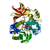 1wooS C: citing same article ( S: Starting model for refinement |
|---|---|
| Similar structure data |


-Links
 PDBj
PDBj- Assembly
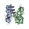
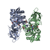
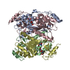
| Deposited unit | 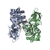
| ||||||||
|---|---|---|---|---|---|---|---|---|---|
| 1 |
| ||||||||
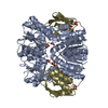
| Unit cell |
|
-Components
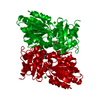
| #1: Protein | Mass: 41886.719 Da / Num. of mol.: 2 Source method: isolated from a genetically manipulated source Details: Gene synthesized with E. coli codon preferences. Plasmid based on pCYB1. Source: (gene. exp.) Candidatus Pelagibacter ubique (bacteria)Strain: HTCC1062 / Gene: dmdA, SAR11_0246 / Plasmid: pABX101 / Production host: Escherichia coli (E. coli) / Strain (production host): Top10F' / References: UniProt: Q4FP21, aminomethyltransferase#2: Chemical | 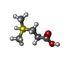  Mass: 136.213 Da / Num. of mol.: 2 / Source method: obtained synthetically / Formula: C5H12O2S Mass: 136.213 Da / Num. of mol.: 2 / Source method: obtained synthetically / Formula: C5H12O2S#3: Chemical | Glycerol  Mass: 92.094 Da / Num. of mol.: 2 / Source method: obtained synthetically / Formula: C3H8O3 Mass: 92.094 Da / Num. of mol.: 2 / Source method: obtained synthetically / Formula: C3H8O3#4: Chemical |   Mass: 22.990 Da / Num. of mol.: 2 / Source method: obtained synthetically / Formula: Na Mass: 22.990 Da / Num. of mol.: 2 / Source method: obtained synthetically / Formula: Na#5: Water | ChemComp-HOH / | Water Mass: 18.015 Da / Num. of mol.: 677 / Source method: isolated from a natural source / Formula: H2O Mass: 18.015 Da / Num. of mol.: 677 / Source method: isolated from a natural source / Formula: H2O |
|---|
-Experimental details
-Experiment
| Experiment | Method: X-RAY DIFFRACTION / Number of used crystals: 1 |
|---|
- Sample preparation
| Crystal | Density Matthews: 2.29 Å3/Da / Density % sol: 46.22 % |
|---|---|
| Crystal grow | Temperature: 298 K / pH: 6.8 Details: 25 mM HEPES, 325 mM NaCl, 20% PEG. PEG increased to 30% for cryo. Soaked with DMSP, pH 6.8, Microbatch under oil, temperature 298K |
-Data collection
| Diffraction | Mean temperature: 100 K |
|---|---|
| Diffraction source | Source: SYNCHROTRON / Site: APS  / Beamline: 22-ID / Wavelength: 0.98 Å / Beamline: 22-ID / Wavelength: 0.98 Å |
| Detector | Type: MARMOSAIC 300 mm CCD / Detector: CCD / Date: Oct 25, 2008 / Details: vertical focusing mirrors |
| Radiation | Monochromator: double crystal Si(111) cooled with lN2 / Protocol: SINGLE WAVELENGTH / Monochromatic (M) / Laue (L): M / Scattering type: x-ray |
| Radiation wavelength | Wavelength: 0.98 Å / Relative weight: 1 |
| Reflection | Resolution: 1.6→50 Å / Num. all: 99295 / Num. obs: 96714 / % possible obs: 97.4 % / Observed criterion σ(F): 0 / Observed criterion σ(I): 0 / Redundancy: 4.5 % / Biso Wilson estimate: 21.2 Å2 / Rsym value: 0.08 / Net I/σ(I): 23.9 |
| Reflection shell | Resolution: 1.6→1.66 Å / Redundancy: 4.6 % / Mean I/σ(I) obs: 10 / Num. unique all: 9909 / Rsym value: 0.151 / % possible all: 99.8 |
- Processing
| Software |
| ||||||||||||||||||||||||||||||||||||||||||||||||||||||||||||||||||||||||||||||||||||||||||
|---|---|---|---|---|---|---|---|---|---|---|---|---|---|---|---|---|---|---|---|---|---|---|---|---|---|---|---|---|---|---|---|---|---|---|---|---|---|---|---|---|---|---|---|---|---|---|---|---|---|---|---|---|---|---|---|---|---|---|---|---|---|---|---|---|---|---|---|---|---|---|---|---|---|---|---|---|---|---|---|---|---|---|---|---|---|---|---|---|---|---|---|
| Refinement | Method to determine structure: MOLECULAR REPLACEMENT Starting model: PDB entries 1WOO, 1V5V Resolution: 1.6→29.2 Å / Cor.coef. Fo:Fc: 0.971 / Cor.coef. Fo:Fc free: 0.949 / SU B: 2.843 / SU ML: 0.047 / Isotropic thermal model: ANISOTROPIC / Cross valid method: THROUGHOUT / ESU R Free: 0.081 / Stereochemistry target values: MAXIMUM LIKELIHOOD / Details: HYDROGENS HAVE BEEN ADDED IN THE RIDING POSITIONS
| ||||||||||||||||||||||||||||||||||||||||||||||||||||||||||||||||||||||||||||||||||||||||||
| Solvent computation | Ion probe radii: 0.8 Å / Shrinkage radii: 0.8 Å / VDW probe radii: 1.4 Å / Solvent model: BABINET MODEL WITH MASK | ||||||||||||||||||||||||||||||||||||||||||||||||||||||||||||||||||||||||||||||||||||||||||
| Displacement parameters | Biso mean: 21.111 Å2
| ||||||||||||||||||||||||||||||||||||||||||||||||||||||||||||||||||||||||||||||||||||||||||
| Refinement step | Cycle: LAST / Resolution: 1.6→29.2 Å
| ||||||||||||||||||||||||||||||||||||||||||||||||||||||||||||||||||||||||||||||||||||||||||
| Refine LS restraints |
| ||||||||||||||||||||||||||||||||||||||||||||||||||||||||||||||||||||||||||||||||||||||||||
| LS refinement shell | Resolution: 1.6→1.642 Å / Total num. of bins used: 20
|
