 Movie
Movie Controller

[English] 日本語
 Yorodumi
- PDB-5cid: Complex of yeast cytochrome c peroxidase (W191G) bound to o-tolui... -
+ Open data

- Basic information
| Entry | Database: PDB / ID: 5cid | |||||||||
|---|---|---|---|---|---|---|---|---|---|---|
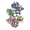
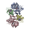
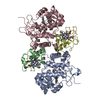
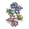
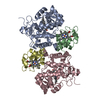
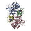
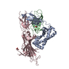
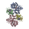
| Title | Complex of yeast cytochrome c peroxidase (W191G) bound to o-toluidine with iso-1 cytochrome c | |||||||||
 Components Components |
| |||||||||
 Keywords Keywords | ELECTRON TRANSPORT/OXIDOREDUCTASE /  electron transfer / heme proteins / electron hopping / multi-step tunneling / photochemistry / ELECTRON TRANSPORT-OXIDOREDUCTASE complex electron transfer / heme proteins / electron hopping / multi-step tunneling / photochemistry / ELECTRON TRANSPORT-OXIDOREDUCTASE complex | |||||||||
| Function / homology |  Function and homology information Function and homology informationRelease of apoptotic factors from the mitochondria / Pyroptosis / Detoxification of Reactive Oxygen Species / Respiratory electron transport / cytochrome-c peroxidase / cytochrome-c peroxidase activity / mitochondrial electron transport, cytochrome c to oxygen / mitochondrial electron transport, ubiquinol to cytochrome c / respirasome / response to reactive oxygen species ...Release of apoptotic factors from the mitochondria / Pyroptosis / Detoxification of Reactive Oxygen Species / Respiratory electron transport / cytochrome-c peroxidase / cytochrome-c peroxidase activity / mitochondrial electron transport, cytochrome c to oxygen / mitochondrial electron transport, ubiquinol to cytochrome c / respirasome / response to reactive oxygen species / hydrogen peroxide catabolic process / peroxidase activity / mitochondrial intermembrane space / cellular response to oxidative stress / electron transfer activity / mitochondrial matrix / heme binding / mitochondrion / metal ion bindingSimilarity search - Function | |||||||||
| Biological species |  Saccharomyces cerevisiae (brewer's yeast) Saccharomyces cerevisiae (brewer's yeast) | |||||||||
| Method | X-RAY DIFFRACTION / SYNCHROTRON / Resolution: 2.763 Å | |||||||||
 Authors Authors | Crane, B.R. / Payne, T.M. | |||||||||
 Citation Citation | Journal: Biochemistry / Year: 2016 Title: Constraints on the Radical Cation Center of Cytochrome c Peroxidase for Electron Transfer from Cytochrome c. Authors: Payne, T.M. / Yee, E.F. / Dzikovski, B. / Crane, B.R. | |||||||||
| History |
|
- Structure visualization
| Structure viewer | Molecule: MolmilJmol/JSmol |
|---|
- Downloads & links
-Download
| PDBx/mmCIF format | 5cid.cif.gz | 171.3 KB | Display | PDBx/mmCIF format |
|---|---|---|---|---|
| PDB format | pdb5cid.ent.gz | 141.8 KB | Display | PDB format |
| PDBx/mmJSON format | 5cid.json.gz | Tree view | PDBx/mmJSON format | |
| Others |  Other downloads Other downloads |
-Validation report
| Arichive directory | https://data.pdbj.org/pub/pdb/validation_reports/ci/5cidftp://data.pdbj.org/pub/pdb/validation_reports/ci/5cid | HTTPS FTP |
|---|
-Related structure data
| Related structure data |  5cibC  5cicC 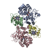 5cieC  5cifC 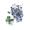 5cigC 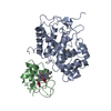 5cihC C: citing same article ( |
|---|---|
| Similar structure data |


-Links
 PDBj
PDBj


- Assembly
| Deposited unit | 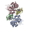
| ||||||||
|---|---|---|---|---|---|---|---|---|---|
| 1 |
| ||||||||
| Unit cell |
|
-Components
| #1: Protein | / CCP Mass: 33396.102 Da / Num. of mol.: 2 / Fragment: W191G Source method: isolated from a genetically manipulated source Source: (gene. exp.) Saccharomyces cerevisiae (strain ATCC 204508 / S288c) (yeast)Strain: ATCC 204508 / S288c / Gene: CCP1, CCP, CPO, YKR066C / Plasmid: ppSUMO / Production host:  Escherichia coli (E. coli) / Strain (production host): BL21 (DE3) / References: UniProt: P00431, cytochrome-c peroxidase Escherichia coli (E. coli) / Strain (production host): BL21 (DE3) / References: UniProt: P00431, cytochrome-c peroxidase#2: Protein | Mass: 12073.835 Da / Num. of mol.: 2 Source method: isolated from a genetically manipulated source Source: (gene. exp.) Saccharomyces cerevisiae (strain ATCC 204508 / S288c) (yeast)Strain: ATCC 204508 / S288c / Gene: CYC1, YJR048W, J1653 / Plasmid: PBTR-1 / Production host: Escherichia coli (E. coli) / Strain (production host): BL21 (DE3) / References: UniProt: P00044#3: Chemical | ChemComp-HEC / Heme C  Mass: 618.503 Da / Num. of mol.: 4 / Source method: obtained synthetically / Formula: C34H34FeN4O4 Mass: 618.503 Da / Num. of mol.: 4 / Source method: obtained synthetically / Formula: C34H34FeN4O4#4: Chemical | O-Toluidine  Mass: 107.153 Da / Num. of mol.: 2 / Source method: obtained synthetically / Formula: C7H9N Mass: 107.153 Da / Num. of mol.: 2 / Source method: obtained synthetically / Formula: C7H9N#5: Water | ChemComp-HOH / | Water Mass: 18.015 Da / Num. of mol.: 9 / Source method: isolated from a natural source / Formula: H2O Mass: 18.015 Da / Num. of mol.: 9 / Source method: isolated from a natural source / Formula: H2O |
|---|
-Experimental details
-Experiment
| Experiment | Method: X-RAY DIFFRACTION / Number of used crystals: 1 |
|---|
- Sample preparation
| Crystal | Density Matthews: 2.38 Å3/Da / Density % sol: 48.27 % |
|---|---|
| Crystal grow | Temperature: 298 K / Method: vapor diffusion, sitting drop / Details: PEG 3350 15-15%, 100 mM NaAcetate, 175 mM NaCl / PH range: 4.8-5.6 |
-Data collection
| Diffraction | Mean temperature: 100 K | ||||||||||||||||||||||||||||||||||||||||||||||||||||||||||||||||||||||||||||||||||||||||||||||||||||||||||||||||||||||||||||||
|---|---|---|---|---|---|---|---|---|---|---|---|---|---|---|---|---|---|---|---|---|---|---|---|---|---|---|---|---|---|---|---|---|---|---|---|---|---|---|---|---|---|---|---|---|---|---|---|---|---|---|---|---|---|---|---|---|---|---|---|---|---|---|---|---|---|---|---|---|---|---|---|---|---|---|---|---|---|---|---|---|---|---|---|---|---|---|---|---|---|---|---|---|---|---|---|---|---|---|---|---|---|---|---|---|---|---|---|---|---|---|---|---|---|---|---|---|---|---|---|---|---|---|---|---|---|---|---|
| Diffraction source | Source: SYNCHROTRON / Site: CHESS  / Beamline: A1 / Wavelength: 0.9778 Å / Beamline: A1 / Wavelength: 0.9778 Å | ||||||||||||||||||||||||||||||||||||||||||||||||||||||||||||||||||||||||||||||||||||||||||||||||||||||||||||||||||||||||||||||
| Detector | Type: ADSC QUANTUM 210 / Detector: CCD / Date: May 23, 2011 | ||||||||||||||||||||||||||||||||||||||||||||||||||||||||||||||||||||||||||||||||||||||||||||||||||||||||||||||||||||||||||||||
| Radiation | Protocol: SINGLE WAVELENGTH / Monochromatic (M) / Laue (L): M / Scattering type: x-ray | ||||||||||||||||||||||||||||||||||||||||||||||||||||||||||||||||||||||||||||||||||||||||||||||||||||||||||||||||||||||||||||||
| Radiation wavelength | Wavelength: 0.9778 Å / Relative weight: 1 | ||||||||||||||||||||||||||||||||||||||||||||||||||||||||||||||||||||||||||||||||||||||||||||||||||||||||||||||||||||||||||||||
| Reflection | Resolution: 2.75→50 Å / Num. obs: 20376 / % possible obs: 93.5 % / Redundancy: 5.4 % / Biso Wilson estimate: 50.57 Å2 / Rmerge(I) obs: 0.092 / Χ2: 1.019 / Net I/av σ(I): 17.182 / Net I/σ(I): 7.7 / Num. measured all: 109129 | ||||||||||||||||||||||||||||||||||||||||||||||||||||||||||||||||||||||||||||||||||||||||||||||||||||||||||||||||||||||||||||||
| Reflection shell | Diffraction-ID: 1 / Rejects: 0
|
- Processing
| Software |
| |||||||||||||||||||||||||||||||||||||||||||||||||||||||||||||||||||||||||||||||||||||||||||||||||||||||||
|---|---|---|---|---|---|---|---|---|---|---|---|---|---|---|---|---|---|---|---|---|---|---|---|---|---|---|---|---|---|---|---|---|---|---|---|---|---|---|---|---|---|---|---|---|---|---|---|---|---|---|---|---|---|---|---|---|---|---|---|---|---|---|---|---|---|---|---|---|---|---|---|---|---|---|---|---|---|---|---|---|---|---|---|---|---|---|---|---|---|---|---|---|---|---|---|---|---|---|---|---|---|---|---|---|---|---|
| Refinement | Resolution: 2.763→42.58 Å / SU ML: 0.37 / Cross valid method: FREE R-VALUE / σ(F): 1.38 / Phase error: 31.52 / Stereochemistry target values: ML
| |||||||||||||||||||||||||||||||||||||||||||||||||||||||||||||||||||||||||||||||||||||||||||||||||||||||||
| Solvent computation | Shrinkage radii: 0.9 Å / VDW probe radii: 1.11 Å / Solvent model: FLAT BULK SOLVENT MODEL | |||||||||||||||||||||||||||||||||||||||||||||||||||||||||||||||||||||||||||||||||||||||||||||||||||||||||
| Displacement parameters | Biso max: 211.29 Å2 / Biso mean: 70.2455 Å2 / Biso min: 23.29 Å2 | |||||||||||||||||||||||||||||||||||||||||||||||||||||||||||||||||||||||||||||||||||||||||||||||||||||||||
| Refinement step | Cycle: final / Resolution: 2.763→42.58 Å
| |||||||||||||||||||||||||||||||||||||||||||||||||||||||||||||||||||||||||||||||||||||||||||||||||||||||||
| Refine LS restraints |
| |||||||||||||||||||||||||||||||||||||||||||||||||||||||||||||||||||||||||||||||||||||||||||||||||||||||||
| LS refinement shell | Refine-ID: X-RAY DIFFRACTION / Total num. of bins used: 14
|
