 Movie
Movie Controller


+ Open data

- Basic information
| Entry | Database: PDB / ID: 5b21 | ||||||
|---|---|---|---|---|---|---|---|
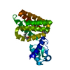
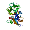
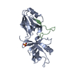
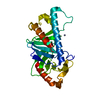
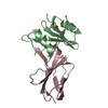
| Title | Dimer structure of murine Nectin-1 D1 | ||||||
 Components Components | murine Nectin-1 D1 | ||||||
 Keywords Keywords |  CELL ADHESION / Cell Adhesion Molecules / CAM / Immunoglobulin-like domains CELL ADHESION / Cell Adhesion Molecules / CAM / Immunoglobulin-like domains | ||||||
| Function / homology |  Function and homology information Function and homology informationNectin/Necl trans heterodimerization / desmosome organization / protein localization to cell junction / Adherens junctions interactions / lens morphogenesis in camera-type eye / growth cone membrane / enamel mineralization / camera-type eye morphogenesis / cell-cell contact zone / virion binding ...Nectin/Necl trans heterodimerization / desmosome organization / protein localization to cell junction / Adherens junctions interactions / lens morphogenesis in camera-type eye / growth cone membrane / enamel mineralization / camera-type eye morphogenesis / cell-cell contact zone / virion binding / heterophilic cell-cell adhesion via plasma membrane cell adhesion molecules / regulation of synapse assembly / apical junction complex / homophilic cell adhesion via plasma membrane adhesion molecules / cell adhesion molecule binding / axon guidance / adherens junction / cell-cell adhesion / retina development in camera-type eye / presynaptic membrane / signaling receptor activity / iron ion transport / carbohydrate binding / membrane => GO:0016020 / neuron projection / symbiont entry into host cell / axon / intracellular membrane-bounded organelle / synapse / dendrite / protein-containing complex binding / virion attachment to host cell / protein homodimerization activity / identical protein binding / plasma membraneSimilarity search - Function | ||||||
| Biological species |  Mus musculus (house mouse) Mus musculus (house mouse) | ||||||
| Method | X-RAY DIFFRACTION / SYNCHROTRON / MOLECULAR REPLACEMENT / Resolution: 2.24 Å | ||||||
 Authors Authors | Sangawa, T. / Takebe, K. / Suzuki, M. | ||||||
 Citation Citation | Journal: To Be Published Title: Dimer structure of murine Nectin-1 D1 Authors: Sangawa, T. / Takebe, K. / Suzuki, M. | ||||||
| History |
|
- Structure visualization
| Structure viewer | Molecule: MolmilJmol/JSmol |
|---|
- Downloads & links
-Download
| PDBx/mmCIF format | 5b21.cif.gz | 104.7 KB | Display | PDBx/mmCIF format |
|---|---|---|---|---|
| PDB format | pdb5b21.ent.gz | 79.9 KB | Display | PDB format |
| PDBx/mmJSON format | 5b21.json.gz | Tree view | PDBx/mmJSON format | |
| Others |  Other downloads Other downloads |
-Validation report
| Arichive directory | https://data.pdbj.org/pub/pdb/validation_reports/b2/5b21ftp://data.pdbj.org/pub/pdb/validation_reports/b2/5b21 | HTTPS FTP |
|---|
-Related structure data
| Related structure data |  3alpS S: Starting model for refinement |
|---|---|
| Similar structure data |


-Links
 PDBj
PDBj


- Assembly
| Deposited unit | 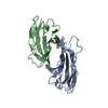
| ||||||||
|---|---|---|---|---|---|---|---|---|---|
| 1 |
| ||||||||
| Unit cell |
|
-Components
| #1: Protein | Mass: 12689.452 Da / Num. of mol.: 2 / Fragment: UNP residues 31-144 Source method: isolated from a genetically manipulated source Source: (gene. exp.) Mus musculus (house mouse) / Gene: Pvrl1, Hvec, Prr1 / Plasmid: pFastBac-1 / Cell line (production host): Sf-9 / Organ (production host): ovary / Production host:   Spodoptera frugiperda (fall armyworm) / References: UniProt: Q9JKF6 Spodoptera frugiperda (fall armyworm) / References: UniProt: Q9JKF6#2: Water | ChemComp-HOH / | Water Mass: 18.015 Da / Num. of mol.: 18 / Source method: isolated from a natural source / Formula: H2O Mass: 18.015 Da / Num. of mol.: 18 / Source method: isolated from a natural source / Formula: H2O |
|---|
-Experimental details
-Experiment
| Experiment | Method: X-RAY DIFFRACTION / Number of used crystals: 1 |
|---|
- Sample preparation
| Crystal | Density Matthews: 2.49 Å3/Da / Density % sol: 50.64 % |
|---|---|
| Crystal grow | Temperature: 293 K / Method: vapor diffusion, hanging drop / Details: 40% PEG3350, 70 mM Na-citrate pH 5.0 / PH range: 5.0-7.5 |
-Data collection
| Diffraction | Mean temperature: 100 K |
|---|---|
| Diffraction source | Source: SYNCHROTRON / Site: SPring-8  / Beamline: BL44XU / Wavelength: 0.9 Å / Beamline: BL44XU / Wavelength: 0.9 Å |
| Detector | Type: BRUKER SMART 6500 / Detector: CCD / Date: Jul 8, 2015 |
| Radiation | Protocol: SINGLE WAVELENGTH / Monochromatic (M) / Laue (L): M / Scattering type: x-ray |
| Radiation wavelength | Wavelength: 0.9 Å / Relative weight: 1 |
| Reflection | Resolution: 2.24→39.16 Å / Num. obs: 12470 / % possible obs: 100 % / Redundancy: 7 % / Rmerge(I) obs: 0.162 / Net I/σ(I): 7.3 |
| Reflection shell | Resolution: 2.238→2.318 Å / Redundancy: 7 % / % possible all: 100 |
- Processing
| Software |
| |||||||||||||||||||||||||||||||||||||||||||||||||||||||||||||||||||||||||||
|---|---|---|---|---|---|---|---|---|---|---|---|---|---|---|---|---|---|---|---|---|---|---|---|---|---|---|---|---|---|---|---|---|---|---|---|---|---|---|---|---|---|---|---|---|---|---|---|---|---|---|---|---|---|---|---|---|---|---|---|---|---|---|---|---|---|---|---|---|---|---|---|---|---|---|---|---|
| Refinement | Method to determine structure: MOLECULAR REPLACEMENT Starting model: 3ALP Resolution: 2.24→39.16 Å / SU ML: 0.4 / Cross valid method: FREE R-VALUE / σ(F): 1.38 / Phase error: 24.77 / Stereochemistry target values: ML
| |||||||||||||||||||||||||||||||||||||||||||||||||||||||||||||||||||||||||||
| Solvent computation | Shrinkage radii: 0.9 Å / VDW probe radii: 1.11 Å / Solvent model: FLAT BULK SOLVENT MODEL | |||||||||||||||||||||||||||||||||||||||||||||||||||||||||||||||||||||||||||
| Refinement step | Cycle: LAST / Resolution: 2.24→39.16 Å
| |||||||||||||||||||||||||||||||||||||||||||||||||||||||||||||||||||||||||||
| Refine LS restraints |
| |||||||||||||||||||||||||||||||||||||||||||||||||||||||||||||||||||||||||||
| LS refinement shell | Refine-ID: X-RAY DIFFRACTION / % reflection obs: 100 %
| |||||||||||||||||||||||||||||||||||||||||||||||||||||||||||||||||||||||||||
| Refinement TLS params. | Method: refined / Refine-ID: X-RAY DIFFRACTION
| |||||||||||||||||||||||||||||||||||||||||||||||||||||||||||||||||||||||||||
| Refinement TLS group |
|
